


Έρευνες
Geriativet cat - Senior Dogs and Cats – a short story
 Abstract
Aging is a natural physiological process that requires increased senior protection. It is associated with progressive changes resulting from dysregulation of homeostasis, greater susceptibility to oxidative stress and lower immunity. Old age in animals is also associated with changes in behavior resulting from the aging of the brain, impaired perception of the sensory organs, etc. Physical and mental activity supported by an appropriate diet and/or supplements allows to delay a number of changes related to aging. The essence of the medical-veterinary approach seems to be the holistic idea of care, supported by the optimization/adjustment of the environment to the needs of the elderly.
Aging is a normal physiological period in the life of an animal for the fnal third of its predicted life. In biological terms, it is associated with progressive changes associated with the deterioration of the functioning of organs, systems, and the entire body. This causes an inability to maintain an endogenous balance (homeostasis), greater exposure to oxidative stress and higher susceptibility to disease, resulting, for example, from reduced immunity, impaired organ function, etc. Old age in animals also means changes in behaviour, caused by degenerative changes in the brain, sensory organs, etc., decreased physical activity (sarcopenia, degenerative changes in joints, etc.) and reduced appetite (1,5).
When the animal becomes a senior
In dogs and cats there are two stages of old age, which can be described as „early” and „late”. In the case of dogs, it is closely related to the size of the dog (breed), in cats, due to the greater uniformity of their bodyweights, there is not so much racial difference (1,5). Simplifed age limits are given in the table below (Tab.1).
Caring for an elderly animal
Animal ageing can also be considered in two aspects: the current problem with senior animals and the prevention or rather delay of the ageing process. Studies in humans indicate that the feeling of age is individual-dependent and strongly associated with physical and mental activity (4,6). There are no contraindications that the above rule of active life and stimulation of the central nervous system could also be applied to animals.
The active life of an older dog is often an extension of his former existence in the environment. This largely depends on the activity and willingness of the owner, but given the working dog parks, you can choose those options for physical entertainment that will suit the animals capabilities. Very important is also the stimulative effect of the environment on the brain cortex of animals, so as to ensure that it is constantly stimulated (7,8). Playing is benefcial for example in the search for delicacies or favorite toys, or other
tasks that require the dog to think about how to do it. It is much more diffcult to activate a cat, due to its nature, considering only 20% of activity during the day and as much as 80% of sleep. Nevertheless, the solution is also a playground with interesting, perhaps natural elements – tree trunks, logs, wooden scratcher, labyrinth, shelves mounted at different heights, interesting tunnels and lockers etc. Owners of houses with a garden, some of which can be adapted to the cat’s „aviary”, have much more possibilities. The possibility of tracking the outside environment is an undeniable beneft that keeps the cat in a good „mental-physical” condition. Unfortunately, we do not know what the cat thinks about sitting on the window or on the veranda of the house, however, from the description of the owners of sick cats, having the possibility to use the „catwalk”. (garden, etc.), we obtain information that they recover relatively faster than those sitting off in the apartments. It seems that an undeniable (and underestimated) beneft is the impact of nature, external environment (greenery, sun, etc.)
Support and stimulation of the central nervous system (CNS) is not possible without proper nutrition. B vitamins are most important in the regeneration processes. However, besides them, protection from free radicals is of great importance. Each cell in the course of evolution has protection mechanisms against reactive oxygen forms. In case of their excessive „exploitation” (high physical exertion, illness of other forms of oxidative stress) or their „ fatigue” progressing with age, may cause problems with maintaining homeostasis and effective protection against oxidation. The solution is then antioxidants, whose main source is the diet or dietary supplements. The best known antioxidant complexes are vitamin E + C, polyphenols, carotenoids, some amino acids (taurine) and others (2,8).
Seniors often have a reduced appetite, resulting from many reasons. This can result in a reduced intake of nutrients. If this is combined with their reduced absorption resulting from digestive restrictions (reduced secretion, enzyme activity, weakened motility, damage to the epithelium of the gastrointestinal tract, change in microflora, etc.), it can
lead to the development of serious nutritional defciencies in the long term. The solution is to introduce supplements containing ingredients that may be potentially defcient in the elderly or for which metabolic needs may be increased. In older cats, the supply of B vitamins and some amino acids such as taurine, methionine or arginine is benefcial. It also supports immunity and protection from the damaging effects of free oxygen radicals. In older dogs, mobility problems, vision impairment or CNS changes often come to the fore, limiting the dog’s contact with its owner and affecting a number of behavioural problems. It is therefore benefcial to introduce supplements to support the functioning of joints, vision or brain work (3,8).
Due to the variety of health problems faced by seniors, the medical and nutritional approach should be guided by the principle of supporting the whole body, rather than focusing exclusively on single organ dysfunctions. According to the above hypothesis, older animals should be adopted to a holistic approach. In highly industrialized countries it is quite diffcult to realize this idea, due to the „civilization rush” and the desire to achieve a quick result of any therapy. Nevertheless, gradual and slow changes with the conscious assumption that the animal won’t be any younger seems to be the right direction to take with the seniors.
dr n. wet. Agnieszka Kurosad
Vet Planet Sp. z o.o.
ul. Brukowa 36/2,
05-092 Łomianki
Neurosupport - Natural sources of selected antioxidants used in a form of dog diet supplements
 Abstract
Healthy organism is in a state of „red-ox” equilibrium, thanks to the cellular protective system that prevents or interrupts the formation and effect of free radicals. That system is supported by antioxidants, contained in the diet or taken in the form of supplements. Their main sources are extracts, dried or other forms of whole plants or their parts: roots, leaves, fruits, flowers, etc. The current nutrition of dogs aimed at natural ingredients also involves the use of antioxidants from their natural sources.
Oxidative stress and protective mechanisms
Oxidative stress is an underlying mechanism of many diseases, including generalized infection of the whole body (sepsis), respiratory and cardiovascular diseases, diabetes
or neurological diseases (8, 23, 25).
Cells, in the course of evolution, became equipped with natural ‘antioxidative’ protection composed of enzymatic mechanisms (superoxide dismutase – SOD, glutathione peroxidase – GPx, catalase – CAT). This system is supported by non-enzymatic low molecular weight antioxidants (vitamin A, C and E) whose main source is diet (23).
The synergy between both mechanisms provides a cell with ‘oxidative stability’ which allows to maintain the body in health. However, maintenance of the ‘liable’ balance is quite diffcult. Therefore potentially each factor which creates imbalance results in triggering the mechanism of cascade: free radicals, oxidation of protein, fat, enzymes, lack of mucous membrane stability, secondary cellular damage and its dysfunctionality. Therefore, the activity of a cellular protection system mainly consists in preventing formation and reaction between reactive radicals and cellular components or inhibiting free radical chain reactions (8).
The foundation of optimum antioxidative protection of the body is correct diet balancing, supplementation with proper antioxidants and effective activity of mitochondrial cofactors (3, 18, 25). They include alpha lipoic acid (LA) and acetylo-L-carnitine (ALCAR) which reduce production of free oxygen radicals in energetic centres of cells – mitochondria. Alpha-lipoic acid plays a role of both a cofactor of a respiratory chain reaction as well as an antioxidant. Whereas acetylo-L-carnitine is a source which contributes
to recovery of depleting L-carnitine reserve and additionally facilitates synthesis of ATP and a process of beta oxidation which takes place in tissues (10, 11, 26). Since in animals which are exposed to a long-term oxidative stress, cellular cofactors are not fully effective, antioxidative protection is enhanced with antioxidants from a diet. Properly formulated complexes are usually used because their accumulative effect is stronger than when they are used individually (29). However, studies of optimal composition of antioxidants, in relation to specifc needs (inhibition of age-related processes, supporting oncological and nephrology therapy etc.) are still in progress. Additional problem related to their effective in vivo activity may result from their varied bioavailability, a limited possibility to reach their target sites and a relatively rapid degradation. Therefore the formulated antioxidant combinations differ between each other with a composition, form or a method of administration in order to make the most of the effects of individual components and the whole complex. The source of the majority of antioxidants, present on the nutritional markets, are natural extracts as well as dried forms of the popular plants, herbs or fruits.
Selected natural sources of antioxidants
and their practical use
One of the most interesting antioxidants of the previous years is curcumin, a very strong antioxidant which belongs to a group of polyphenols. Its main source is a dried root of Curcuma longa. Curcumin, which is related to as a ‘herbal aspirin or ‘herbal cortisone’ is used in ayurvedic medicine to alleviate inflammation as well as to treat infectious and immune-mediated diseases (13). Curcumin signifcantly reduces peroxidase of lipids, regulates antioxidant enzymes and ‘radical catchers’. In redox reactions it plays a role of an electron donor. It also shows an anti-inflammatory activity which, when combined with antioxidative function, is used to support many therapies including those of the gastrointestinal system (liver and pancreas), skin, joints, nervous system or cancer. Curcumin is also used in an antioxidative model of supporting therapy
of degenerative diseases with etiopathogenesis attributed to destructive effects of free radicals. Its application in human medicine in therapy of Alzheimer and Parkinson diseases or even depression is widely described (22).
The biggest advantage of curcumin is its good tolerance by the body and lack of toxicity. The main limitation however is its poor bioavailability resulting from a limited water solubility, poor stability, rapid pass effect and liver metabolism. Curcuminoids are also relatively rapidly excreted from the body, mainly with faeces. An estimated
1% of active substances enters into the circulatory system. Therefore, many studies were performed in order to increase its bioavailability. Heating of the solution of curcumin increases its bioavailability 12 times. The same effect is observed in case of consumption of curcumin with fat (in particular with coconut oil) or a pepper (piperine).
Useful combinations of curcumin, in a form of phospholipid, polysaccharide, liposome complexes or micelles and nanomolecules are used. It facilitates its penetration across the brain-blood barrier ensuring an effective activity in its target site. Therefore, nanocurcumin whose size does not exceed 100 nm is used in supporting patients with disorders of a central nervous system (22). So far optimal dosing of curcumin and curcuminoids in humans was not specifed. According to recommendations of the European Food Safety Authority (EFSA), a daily dose for people is 3 mg/kg of body weight. In case of healthy persons who want to use curcumin as an element of health promotion and prevention, there are no limitations, except for the mentioned dose which should not be exceeded. Pregnant woman are an exception because curcumin, when taken in excessive doses, may cause uterine contractions leading to abortions.
In compliance with EU Regulations No 1831/2003, in animals antioxidants exclusively from herbal extracts, dried or other forms which are approved in the European Union register of feed additives may be used. The dose however usually depends on the intended use, composition and the content of all the substances in the supplement and its form (24, 25).
Catechins from green tea leaves are very interesting and natural antioxidants and they include: epigallocatechin gallusan (EGCG), epicatechin (EC), epicatechin-3-gallate, (ECG) and epigallocatechin (EGC). Apart from green tea, they also may be found in rapes, coffee grains, apples or citrus fruits. Tea also includes other biologically active
ingredients such as flavanols (e.g. chempherol, quercetin, myricetin and their glycosides) natural caffeine (2.5-4.5%), vitamin C (90 mg/100 g of dry matter) which becomes degraded in the process of drying as well as vitamins B1, B2, PP, A, K and minerals (potassium, calcium, magnesium, aluminium, manganese, iron etc.) (2). However, the main ingredient, which is commercially used due to its antioxidant potential, are catechins. They lead to the increase of the activity of main antioxidant enzymes (catalase, superoxide dismutase, quinone reductase, S-transferase, reductase and glutathione peroxidase) as well as regeneration and inhibition of oxygenation of low molecular weight antioxidants (vitamin C and E, glutathione or beta-carotene). Both of these mechanisms contribute to the enhancement of the antioxidant potential of the whole body. However,
its effcacy is strictly related to the cellular antioxidant capacity and in case of acquiring optimum concentration, further improvement is not observed (21). Polyphenols indirectly also affect inhibition of pro-oxidant enzyme activity (lipoxygenases, nitric oxide synthase, cyclooxygenase, xanthine oxidase, myeloperoxidase etc.) showing a destructive effect on DNA and body proteins (21). Ad ditionally, they also show an ability to chelate transition metal ions (iron and copper).
Effective and strong activity (25-100 times more potent than the activity of vitamin C or E) of catechins results from a chemical structure. Therefore they are readily used to support therapy of diseases with various underlying mechanisms (cancer and metabolic diseases: diabetes, obesity, vascular diseases, chronic organ diseases: kidney and liver disorders etc., degenerative (nervous system) or infectious (viral and bacterial) diseases (4, 27). Likewise curcumin, they are usually formulated into complexes. Absorption and pharmacokinetics of catechins is quite precisely described on the basis of a laboratory animal model and beagles. Good absorption, after oral administration of about 200 mg of green tea extract and fairly sustained concentration of metabolites for a long time prove that there is high potential for their use in practice (5). Nevertheless, subsequent studies showed presence of necrotic changes in the cells of liver, kidney and the gastrointestinal tract in dogs. More severe damages were observed in starving animals. Yet it does not fully explain the mechanism of developing changes (28). Similar observations were made in mice confrming dose-dependent toxicity (14). An example of green tea extract shows that a component with potentially favourable effect, due to its potency and long-lasting effect of contained antioxidants, must be carefully dosed to prevent adverse changes.
Another interesting example of a source rich in antioxidants is Malphigia glabra (acerola) fruit extract. West Indian cherry, also called a wild crepemyrtle or Barbarados cherry naturally occurs in South, Central and North America as well as in Madagascar. The source of antioxidants are its fruits with juicy and intensely sour flesh. Acerola is a source of 17 various carotenoids including B-carotene which is present at the highest concentration (up to 75%). Their total concentration in fruits varies from 371 to 1881 mg /100 g (6). Vitamin C is also present in high amount (from 1000 to 45000 mg in 100 g of fresh fruits) but its concentration depends on the stage of development (it is the highest in unripe fruits and then during ripening it becomes reduced as a result of biochemical oxygenation) (15). Additionally, acerola fruits also contain: sugar (glucose, fructose), amino acids (aspartic acid, alanine, proline, serine, GABA), pigments (anthocyanins), minerals (potassium, iron, calcium, phosphorus) and B vitamins (B1, B2, PP) (17). Antioxidant activity of its ingredients is used to support therapy of metabolic diseases such as diabetes (mainly type II), obesity, skin diseases, nervous system disorders (atherosclerosis, Parkinson and Alzheimer diseases in humans) and as a prevention of ageing and weakening of the body or other consequences of an oxidative stress. The antioxidative effect of vitamin C is usually used to formulate supplements for animals.
Fruits and leaves of Vitis vinifera contain: fruit acids, minerals, tannins, anthocyanins, flavonoids, stilbenes (resveratrol), wax, vitamins, procyanidins, pectins, polysaccharides, aromatic substances and carotenoids. Vitis vinifera extract is the most commonly used and apart from antioxidative activity, it also shows antibacterial, anti-inflammatory and antihistaminic effect. Thus it is used to support therapy of skin, liver, heart, central nervous system diseases as well as in treatment of cancer and other conditions (19).
A natural source of antioxidants also may be flower petals e.g. Tagetes erecta extract. Tagetes naturally occurs in Central and South America and it was brought to Europe in 1573. Its flower extract contains about 27% of various carotenoids, including xanthophyll esters (86.1%), cryptoxanthins (1.5%) and beta carotenes (0.4%). Their total value in a dry flower matter is 17.8 g/kg. Lutein and zeaxanthin are the most important components in terms of their use in supplements which improve health. Their activity is focused on the target site in which they accumulate in the body – the macula lutea of the eye. They are also effective when used to support therapy of gastrointestinal inflammatory diseases, mainly ulcerative colitis (16).
The above list of the sources of active substances shows their diversity. However, the composition, dose or fnal form of the supplement are modifed by appropriate elective researches. A pilot study of a combined formulation with vitamin E may serve as an example. The product was used in groups of dogs with various systemic diseases. The study showed lack of signifcant differences between a supplemented and non-supplemented animals in terms of oxidative status and improvement of the clinical condition. Nevertheless, the authors make a reservation that the study had numerous limitations, including wide divergence between disease entities (from infectious to neurological disorders) and a limited duration of the experiment (30 days) which may have had an impact on the outcome (9).
Significantly better results were achieved after using antioxidants in the study which was focused on brain ageing and was based on a canine model (7, 12, 20). With age we observe an increasing lipid peroxidation, protein oxygenation and reduction of the level of endogenous antioxidants which are free radical catchers (12). We do not know whether deposition of amyloid itself or a damage secondary to oxidation generate primary pathological lesions during the brain ageing. However, we observed that antioxidants supplementation of elderly dogs diet for 6 months led to significantly better results in task tests when compared to non-supplemented group of seniors (20). Additionally in the group which was supplemented with antioxidants, stronger antioxidant activity, reduction of protein damage caused by free radicals and limitation of amyloid deposition were found. A study which was performed by Dodd et al in the group of elderly dogs (above 7 years of age) living with their owners was also very important (7). Dog owners were provided with a compound supplement containing a complex of antioxidants: vitamin E, vitamin C, docosahexaenoic acid (DHA), eicosapentaenoic acid (EPA), lipoic acid, L-carnitine as well as dried fruits and vegetables, which was used each day for 2 months. Afterwards, dogs behaviour in terms of disorientation, sleep disorder, daily activity and relationship with a man as well as active training at home were evaluated. In each of those categories they reported a significant improvement of the results when compared to the non-supplemented group (7). A significant observation was also a relatively rapid effect which was visible 2 months after the supplementation. The above studies showed that introduction of antioxidants into diet may prevent changes in animal behaviour resulting from ageing and probably development of neuropathological lesions secondary to oxidative stress. They also prove that the expected effect of their activity may be visible after a relatively short time of their supplementation.
In terms of reducing damages and supporting neurogenesis, the effect of other additives, in particular of B vitamins, folic acid, polyunsatu rated fatty acids etc. should be men tioned too. Boldrini et al. 2018 pre sented very promising studies which showed so called ‘persistent adult neurogenesis’ in healthy people who were in eighth decade of life
and di did not have any cognitive or neuropsychiatric disorders (1). Therefore it may be suspected that by maintaining the body in good health thanks to a balanced diet, supplements and enrichment of the environment, we can benefit from ‘neurons’ much longer that it was previously suspected. The key how ever is homeostasis. It may be also
suspected that the same applies to our animals.
Prof. dr hab. Michał Jank
Zakład Farmakologii i Toksykologii
Instytut Medycyny Weterynaryjnej
SGGW w Warszawie
ul. Ciszewskiego 8,
02-786 Warszawa
CardioVet - To support or not to support – supplements in dogs with advanced heart failure
 Magdalena Garncarz, DVM PhD, Marta Parzeniecka-Jaworska DVM PhD
Department of Pathology and Veterinary Diagnostics, Faculty of Veterinary Medicine,
Warsaw University of Life Sciences – SGGW, Poland
INTRODUCTION
Supplementary therapy for patients with heart diseases has been in use for a long time in different forms. Some of the products available commercially include L-carnitine, a substance necessary for correct metabolism of fatty acids and energy production; taurine that plays a crucial role in normal function of the heart; Q10 coenzyme (ubiquinone), important in energy metabolism of the heart; and vitamin E that – just like Q 10 coenzyme or taurine – has got a strong antioxidative properties. In dogs with different stages of heart insufciency it is difcult to compare the effect of action of these substances themselves without resorting to standard therapy, that is diuretics, inodilators, inhibitors of angiotensin converting enzymes, and antiarrhythmic drugs. Therefore the authors attempted at evaluating how dogs with heart diseases of different stages feel.
MATERIALS AND METHODS
The study was performed on 36 dogs of different breeds, aged from 36 to 222 months, 10 females, 26 males. Majority of dogs were diagnosed with chronic mitral valve disease (CMVD, 25 dogs), while others with dilated cardiomyopathy (DCM, 11 dogs). All dogs had transthoracic echocardiographic test performed. Moreover, x-ray was performed to evaluate the presence of left sided congestive heart failure. The stage of heart insufciency was determined based on accepted standards, following classifcation of International Small Animal Cardiac Health Council (ISACHC). According to this classifcation, dogs with CMVD or DCM were qualifed as class 1 (asymptomatic, n = 9), class 2 (mild to moderate heart failure, n = 16) or class 3 (advanced heart failure, n = 11). On the day of the visit, dogs classifed as ISACHC 2 and 3 received a supplement for dogs with heart failure to complement standard therapy. The composition of the supplement includes: L-Carnitine tartrate 500 mg, taurine 200 mg, Q10 coenzyme 10 mg and vitamin E 60 IU/pill (CardioVet, VetExpert). The product was administered according to producer’s recommendations. After a month, during the follow-up visit, the owners were asked to describe their observations about how the dogs felt, focusing on their effort tolerance during walks, respiratory symptoms (tachypnoea) and coughing.
RESULTS
Out of nine dogs classifed as ISACHC 1, owners of three patients observed improved ftness, describing their dogs as being more lively after administration of the supplement, as compared to the period prior to administration (33.3% of ISACHC 1 dogs, 8.3% of all studied dogs). According to the owners, in 12 out of 16 ISACHC 2 dogs effort tolerance improved – the owners described their dogs as more lively (75% of ISACHC 2 dogs, 33.3% of all studied dogs). This group included eight dogs with CMVD and four dogs with DCM. As regards 11 dogs with the advanced heart failure, namely ISACHC class 3, in nine patients effort tolerance improved (81.8% of ISACHC 3
dogs, 25% of all studied dogs). This group included two out of three dogs with DCM and 7 out of 8 dogs with CMVD. In two dogs (one with CMVD, one with DCM) cough frequency decreased (18.2% of ISACHC 3 dogs, 5.6% of all studied dogs).
SUMMARY
The study showed favorable effect of the analysed product on sick animals with advanced heart failure. The studied product led to improved effort tolerance in majority of dogs, although the evaluation of the effort tolerance was based on subjective impressions of the owners. The owners did not observe adverse effects of the product. The pills were well tolerated, with no adverse effects. Only rarely the problem was the size of the pill. In small breeds it is necessary to crash the pill and administer it mixed with food or water. This study shows good tolerance and effects of the complex product – CardioVet, particularly in dogs with advanced heart failure.
Hepatiale Forte - The effectiveness of preparation Hepatiale Forte in the treatment of the hepatic disorders in dogs.
 The liver is the greatest and the most solid organ in the body, situated strategically between the digestive system and the centre of the circulatory system. It is well vascularized, veins gathering blood from the abdominal cavity and arteries of aortal branches flow into the organ.
The liver is “the central point” of metabolism. Synthesis, detoxification, biotransformation,
haemopoiesis, accumulation and secretion of many chemical compounds and substances take place here.
Antibodies are created here, the liver takes part in the mechanisms of regulation of water-electrolyte
equilibrium, thermoregulation and circulation. Many of these functions and regulations occur only in the
liver. It constitutes, as an organ, around 3.4 % of the body mass of the adult dog, in the young this
percentage is higher(1). The basic structural elements of the liver are hepatic cells- hepatocytes. They are
big, multiwall (eight- or more) cells, sometimes multinuclear, with high metabolic activity. Their
important structural part is the cell membrane, containing receptors for many hormones, plasmatic
proteins and glycoproteins. This unique surface contains specific antigens, typical only for the liver,
lipoproteins reacting to immunological damage in chronic hepatopathies in humans(2).
The liver is an organ extremely easily adapting to changing work conditions of the body. Also high
functional reserve (around 65%) and the ability to regeneration cause, that development of the clinical
symptoms of hepatopathy usually means a serious damage to the liver. As it was mentioned earlier, the
liver influences functioning of the entire body (and vice versa), thus symptoms of the liver diseases are
very various and, sometimes, not connected with the organ in itself. The symptoms of the digestive
system are of prime importance- lack of apetite, vomiting , diarrhoea, constipation, afterwards weight
loss. In the hepatic failure symptoms relating to protein production join to already mentioned- coagulation
disorders with predisposition to bleeding, haemopoiesis disturbances with increasing anaemia. Frequently
jaundice is considered the main symptom of liver disorders, rarely symptoms of polyuria /polydipsia and
neurological disturbances are considered hepatogenic. The symptomatology of liver diseases is then very
abundant, but it lacks typical and distinctive symptoms. Hepatopathies can progress also
asymptomatically. Thus sometimes the diagnose of the liver disease is accidental, for example by making
geriatric profile or the medical examination preceding general anaesthesia (5,10).
In the broad context liver damaging agents can be divided into inflammatory and non-inflammatory.
The first ones are viruses, sometimes liver specific (for example adenovirus I), bacteria, rickettsiae, fungi,
protozoa, parasites. Non-infectious agents are connected with metabolic disorders (for example IBD,
pancreatitis), autoimmunization (haemolytic anaemia), heart diseases (cardiomyopathies) and exposure to
toxins (copper, zinc, algae), medications (phenobarbital, ketoconazole) and biological substances
(aflatoxins). Pathogenesis of hepatocytes degeneration, leading to their necrosis and death is multifactorial, complicated and not completely clear. One points out primary and secondary causes, among them is chaemia and anoxia of tissues, free radicals activity and oxidative injuries, lack of essential intracellular structural elements, intracellular production of toxins, hepatocytes damage causing toxins incorporation into cellular proteins, RNA and DNA of hepatocyte, cholestatic disorders, endotoxins, bacteria, viruses, parasites, immunological mechanisms, the cell membrane injuries and oxidation of fats forming the cell membrane of hepatocyte. Last two mechanisms are particularly important factors inducing hepatocyte disintegration and they can lead secondarily to biochemical and immunological disorders.
The cell losses enzymes, coenzymes, electrolytes.
The cell membrane damage enables access of calcium ions and other electrolytes to the cell and can
lead to irreversible intracellular changes, with possible death of the cell. Injuries of the cell membranes of
the hepatocytes are the best documented as the cause and form of the liver necrosis (3). Attempts of
“reinforcement“ and “repair” of the damaged cell membranes of hepatocytes are based on the use of
phospholipids as the main cell membrane components. Degradation of phospholipids is then an early
effect of activity of majority hepatotoxins and mechanisms damaging the liver. Trauma to the cell
membranes is caused by the disorder of activation of phospholipase A and C, being calcium ions
dependent. The enzymes are essential to creation of information carriers using phosphatidylinositol, that
is an important mechanism of intercellular signalling (2,3). Polyunsaturated phosphatidylcholine was
used as the first phospholipid in trial with humans having active chronic hepatitis and alcoholic
hepatonecrosis (3). Myo-inositol- the main component of phophsatidylocholine is counted in vitamins B
group, it is a lipotropic constituent of the cell membranes and lipoproteins.
It modulates activity of the important cell membrane enzymes for example Na/K ATP-ase, acts as a
mediator in the transmembrane cellular signalling by influencing on protein kinase C. It influences on
intracellular phosphorylation of calcium and proteins.
Phosphatidylocholine production is lowered in the liver diseases. It is thought, that its application in
the treatment of humans having active chronic hepatitis modifies immunological liver damages, what
probably results in restoring signalling function of the cell membranes (3). Phospholipids are esters of
cholinophosphoric acid and unsaturated fatty acids (linoleic, linolenic, oleic). They build themselves in
the cell membrane and in the cytoplasmic reticulum of hepatocytes, filling defects caused by damages
(diseases). It results in faster regeneration of the injured cells and restores their normal function. Thanks
to them the actions of membrane receptors, membrane enzymatic systems and active and passive
transport improve. Phospholipids are also essential for differentiation and proliferation of hepatocytes.
They inhibit processes of hepatic tissue fibrosis by lowering collagen production and increasing
collagenase activity- the enzyme degrading collagen. They play the important part in fats digestion and
vitamins absorption.
Based on above information it was decided to test the effectiveness of phospholipids in the liver diseases
of dogs. They appear relatively frequently in these animals; as mentioned above, often high activity of the
liver enzymes can be the only symptom. Because hepatopathies are treated using many medicaments, this
particular study is restricted to the cases, where the only used preparation was, containing phospholipids,
Hepatiale Forte.
Material and methods
In the trial preparations Hepatiale Forte and Hepatiale Forte Large Breed (+25 kg) by VetExpert
manufacturer were tested, they contain respectively 150 and 275 mg of soya phospholipids (containing
phosphatidylcholine), and 150 and 275 mg of ornithine in the form of L-ornithine L-aspartate. The drug
was given one time a day in dose of 1 tablet/ 15 kg (Hepatiale Forte Large Breed – 1 tablet/25 kg). The
observations were conducted in 22 dogs of the different sex, age and race, the patients of the Veterinary
Polyclinic of the Veterinary Medicine Department at the Warmian-Masurian University in Olsztyn. The
animals were qualified for the trial provided that they had high activity of the hepatic enzymes.
Eight animals were qualified for the test only based on high activity of the liver enzymes detected in
geriatric examinations and tests preceding general anaesthesia. Hepatiale Forte was given to 9 dogs as a
hepatoprotector after long term glycocorticosteroids treatment (3 cases of immunohaemolytic anaemia
following anaplasmosis), glycocorticosteroids and antibiotics treatment (2 cases of idiopathic profound
dermatitis GS), antibiotics and drugs for example antiparasitic (4 cases of general mange). Hepatic
failure was detected in 5 dogs, inflammatory (4 dogs) and cholestatic (1 dog). The medicament was
administered to all the animals for two months. Cases of the liver failure were diagnosed not only basedon activity of the hepatic enzymes and the clinical examinations. They were confirmed by other methods,
which are not the subject of this study.
Blood test (red cell count Erys, white cell count Lkcs, haemtocrit value Ht, haemoglobin Hb, platelet
count PLT) and serum biochemical tests (alanine aminotransferase activity ALT, aspartate
aminotransferase activity AST, alkaline phosphatase activity ALP, total protein level BC, albumins level
ALB, total bilirubin level BIL and urea level UREA)were performed in all the animals. The examinations
took place on day 0- before the drug administration and on day 30th and 60th of the application of
Hepatiale Forte. Before the observation 17 animals had not had liver disease symptoms. The patients
suffering from hepatic failure had had the digestive system disorders- lack of apetite, vomiting, diarrhoea
and general symptoms in the form of weakness, glumness; one dog had been diagnosed with the jaundice.
Hepatiale Forte was given to these animals after the clinical improvement and following appetite return.
Results
Any side effects were not diagnosed in 17 animals either before, or during use, or after finishing
Hepatiale Forte, containing phospholipids, administration. The animals having hepatic failure were
treated with multidrug therapy; as the clinical condition improved, during and after Hepatiale Forte use,
neither symptoms of recurrence of the disease nor other clinical signs were observed. During entire
observation time the animals had a constant water access, were fed with the standard complete volume
fodder. During the experiment they were not given drugs against internal parasites. Prophylaxis against
ticks was used. The preparation Hepatiale Forte was readily eaten by the dogs.
Changes in the erythrocytes of all the dogs were not noted in the haematological tests before (day “0”),
during (day “30th”) and after (day 60th). The value of Erys, Ht, HB and PLT were within the normal range
in the patients having only high activity of the liver enzymes and also in the dogs suffering from the
hepatic failure. Differences were observed within the scope of leukocytes Lkcs. Increase of the leukocytes
Lkcs in the patients treated with potentially hepatotoxic drugs (glycocorticosteroids, antibiotics,
antiparasitic) was mediocre (12.2-19.2x 10⁹ /l), but significant increase was noted in patients having liver
diseases- 38.6-60.3x 10⁹ /l. In all the patients increase of the activity of hepatic enzymes was observed
ALT, AST, ALP, at the same time the greatest one occurred in the patients having the clinical symptoms
and they were respectively: ALT 732-1050 IU/L, AST 205-822 IU/L, ALP 815-2341 IU/L. In the rest of
the dogs the increase was mediocre: ALT 93-533 IU/L, AST 56-305 IU/L, ALP 83-506 IU/L. Total
protein level and albumin level were within the normal range in the “symptomless” dogs and amounted to
BC 43-84 g/l, ALB 28-49 g/l. In the patients having hepatic diseases total protein level BC 23-49 g/l and
albumin ALB 12-22 g/l were lowered. Total bilirubin and urea levels were also maintained within
physiological norms in the dogs not having clinical symptoms and amounted to BIL 0.3- 3.1 µmol/l,
UREA 3.8- 7.11 mmol/l. It was noted that in the animals ill from hepatic diseases total bilirubin level
increased BIL 3.4- 8.2 µmol/l and urea level dropped UREA 1.3-3.0 mmol/l.
In the second test (after 30 days of the use of Hepatiale Forte) the examined parameters tended to
normalization. Leukocytes decrease in the animals not having hepatic symptoms was observed 8.5-19.2x
109 /l and in the ill ones 20.3- 43.5x 10⁹ /l. Also the activity of hepatic enzymes declined in both groups of
animals in the scope of biochemical tests, respectively: ALT 46- 257 IU/L and 301-602 IU/L, AST 28-
100 IU/L and 93- 405 IU/ L, ALP 83- 260 IU/L and 515- 1003 IU/L. Total protein and albumin levels
stayed within the normal physiological range in the “symptomless” group: BC 55- 73 g/l, ALB 29- 41 g/l.
They increased in the “symptomatic” group: BC 31- 55 g/l, ALB 18- 32 g/l. In the group of dogs having
hepatic diseases symptoms total bilirubin level dropped BIL 3.0- 5.4 µmol/l and urea level increased
UREA 1.5- 3.6 mmol /l. In the patients not having clinical symptoms deviation from the physiological
norms for total bilirubin and urea was not noted BIL 0.4- 2.8 µmol/l and UREA 4.0- 6.9 mmol/l.
In the third test, after 60 days of Hepatiale Forte use, further stabilization and normalization of the
parameters were noted. In the group of “symptomless” dogs white blood cell count Lkcs was 8.7- 14.3x
109 /l, activity of ALT 33- 163 IU/L, AST 20- 41 IU/L, ALP 88- 201 IU/L. Levels of total protein BC 56-
75 g/l, albumin ALB 33- 42 g/l, total bilirubin BIL 0.4- 2.1 µmol/l and urea UREA 4.2- 7.5 mmol/l were
also within the scope of the physiological norms. In the group of animals having the symptoms of hepatic
diseases the parameters were also normalized. White blood cell count Lkcs declined to 11.3- 28.3x 10⁹ /l,
activity of ALT 93- 350 IU/L, AST 40- 200 IU/L, ALP 202- 750 IU/L. Levels of total protein, albumin
and and urea increased respectively BC 44- 69 g/l, ALB 21- 40 g/l, UREA 2.8- 4.2 mmol/l. Further
decrease in total bilirubin level was observed 2.8- 4.0 µmol/l.
The values of the particular parameters were shown in the table 1, 2, and 3. For the better presentation of
normalization tendencies and changes within the examined parameters after Hepatiale Forte use one
decided to present the individual patients, without calculating the average values of the parameters.
Discussion
One of the means, used as an auxiliary in the treatment of the liver diseases, especially chronic, are
medications containing phospholipids, coming from soya seeds or salmon eggs. Phospholipids are the
basic component of the cell membranes of all the living creatures- they form the semiliquid matrix, with
the embedded proteins and lipids flowing there. Phosphatidylcholine, constituting about 40% of all
phospholipids of the cell membrane, is one of the most important components providing correct fluidity
and biophysical properties of this cellular structure, thus it has fundamental significance for the proper
cell function. It is also an important element of the blood lipoproteins and the bile. It takes part in the
proper function of the digestive system and the lungs because it contributes to the functional protective
film.
In the practice of the clinical human medicine particularly useful are the medications containing fractions
of phosphatidylcholine, that are abundant in essential polyunsaturated fatty acids. They are characterized
by the very high- about 90%- bioavailability, essential fatty acids are additionally used to produce some
anti-inflammatory eicosanoids. The main indications for their use in humans are all kinds of the liver
damage; they act beneficially also in the cases of cholelithiasis and functional disorders of the biliary
tract. Phospholipids activity is beneficial in curing chronic liver diseases of the different etiology,
including damages caused by alcohol, drugs, toxins. They are used as an auxiliary mean in the treatment
of the infectious jaundice and in the interferon therapy for the liver (6, 7, 13).
Research on ways of the application of phospholipids in the medicine has been carried out since the
1980s. The animals, rats as well as dogs or chimpanzees, serve as the models for active chronic hepatitis
in humans. Also hepatoprotective and modulating activity of phospholipids in patients having alcoholic
cirrhosis of the liver was studied. Tarashi and co-authors found the increase of the membrane tolerance to
ethanol following phospholipids application in rats (12). Waring and co-authors confirmed that the
adaptation changes in the structure of phospholipids lead to the structural changes, that result in the
growth of the mitochondrial membranes resistance to the alcohol-induced damage (14). Phospholipids
protect the hepatocytes and the mitochondrial membranes by increasing their “fluidity”, the cells
proliferation and the incorporation of the enzymes metabolizing drugs into cytoplasmic reticulum (8). It
may explain normalization of the biochemical parameters in dogs treated for a long time with
glycocorticosteroids, antibiotics and antiparasitic drugs, after use of Hepatiale Forte containing
phospholipids.
The application of phospholipids in hepatic disorders appearing during the total parenteral nutrition was
also studied. It is a well-known fact, that the total parenteral nutrition causes hepatocytes damage and the
increase of the hepatic enzymes activity. In the experiment studied group was given phospholipids in doseof 50 mg intravenously every 6 hours for two weeks. Control group was not given the hepatoprotectors.
Statistically significant growth of the ALT, AST, GGT activity in control group was noted in 7th day as
well as in 14th day of the experiment. In the patients taking phospholipids statistically insignificant
increase in ALT activity after 14 days of phospholipids use was observed, without growth in GGT and
ALP activity (9).
Hepatoprotective phospholipids effect is also confirmed by this, the author`s own, study- decline of the
hepatic enzymes activity to the normal range or, in the patients suffering from the liver diseases, a
constant downward trend in their activity (Tab. 1, 2, 3)

Other researchers also confirm beneficial phospholipids effect, paying attention not only to favourable
impact on the liver. Their positive influence on the blood vessels in the arteriosclerosis, ischaemic heart
disease, cardiac infarct, some disorders of the digestive system is underlined thanks to their contribution
to the structure of all the cell membranes of the body (6, 11). According to the last reports, in in vitro
studies and in experiments on animals, their effects were confirmed: antioxidative, anti-inflammatory,
antifibrotic, modulating cell apoptosis, regenerative, reparative and protective for the cell membranes, as
well as influencing on receptors and intercellular signaling and regulating fat metabolism in response to
the damaging activity of toxins and drugs. As a result of the trials conducted in Europe and Asia the
improvement of the clinical, biochemical, imaging and histological parameters was pronounced in the
cases of fatty degeneration of the liver, drug-induced intoxication, and, auxiliary, in viral hepatic diseases
and hepatic coma (7). Normalization of the biochemical parameters in the conducted experiment also
supports these trials.
Most of the researchers did not observe any side effects after administration of phospholipids (4, 7, 9).
Sometimes mild disturbances of the digestive system are mentioned. In author`s own studies the side
effects of use of Hepatiale Forte in dogs were not observed. It exists a certain disagreement in relation to
the taken dose of phospholipids. For the humans it does not exceed 3g/ day. Lata and co-workers used
successfully in their trials dose of 200 mg/ day per an adult (4 x 50 mg) (9). Experimental rats took dose
of 100 mg/ kg, chimpanzees- 4.1 mg/ kg of diet. Doses for the dogs are extrapolated from the human
medicine (2, 4). High content of phospholipids in Hepatiale Forte preparation draws attention- 150 mg in
the tablet per 15 kg of the body mass and 275 mg in the tablet of Hepatiale Forte Large Breed (+25 kg).
Taking into account the positive therapeutic effect demonstrated in this study, the dose seems to be
enough to protect canine liver. However in relation to not established border dose, the trials on the
effectiveness of higher phospholipids dose will be carried out in the future.
Part of the researchers pays attention to antifibrotic phospholipids activity and uses them along with
azathioprine and glycocorticosteroids. It is thought, that they enhance the stabilizing effect of
prednisolone on the cell membranes, without decreasing its growth inhibiting activity. They reduce,
however, fat accumulation and hipoproteinaemia, related to prednisolone activity (3, 13).
Decline and normalization of the activity of hepatic enzymes, increase in total protein, albumin, urea,
decrease in bilirubin when the only used agent was Hepatiale Forte can prove phospholipids usefulness in
the treatment of drug-induced liver damages and hepatopathies demonstrating subclinical and clinical
symptoms. The phospholipids dose, in view of the achieved results, is suitable for the effective treatment
of dogs. Application of the drug containing phospholipids and ornithine seems to be effective, all the
more so because phospholipids partly stabilize the hepatocyte from the “outside”, while ornithine acts
from the “inside” of the cell, by regulation of the urea cycle and by conversing ammonia, coming from
the amino acids breakdown, into urea, decreasing its toxicity. High urea level in dogs having the liver
diseases can be taken as evidence of it.
Complete normalization of the hepatic enzymes activity was not observed in the dogs suffering from the
hepatopathies after 60 days of Hepatiale Forte use. Serious hepatic damages may require longer time of
phospholipids treatment. These patients have been still given Hepatiale Forte, demonstrating lack of side
effects, clinical symptoms of hepatic diseases and further improvement of the liver tests.
Based on the conducted observations, it can be stated, that the drugs Hepatiale Forte and Hepatiale Forte
Large Breed (+25 kg), containing phospholipids and ornithine, are useful and effective in the treatment of
the liver diseases, demonstrating clinical and subclinical course, and in drug-induced hepatic reactions in
dogs. It can be used as the adjunctive and complementary treatment, and also as the hepatoprotective
agent after long lasting pharmacotherapies.
Literature:
1.Center S.A., Strombeck D.R.: Liver: Normal Structure and Function. W: Grant Guliford W.,
Center S.A., Strombeck D.R., Williams D.A., Meyer D.J. eds. Strombeck´s Small Animal
Gastroenterology. 3. Ed Philadelphia, W.B. Saunders Company, 540 – 552
2. Center S.A.: Pathophysiology of Liver Disease: Normal and Abnormal Function. W: Grant
Guliford W., Center S.A., Strombeck D.R., Williams D.A., Meyer D.J. eds. Strombeck´s Small
Animal Gastroenterology. 3. Ed Philadelphia, W.B. Saunders Company, 553 – 632
3. Center S.A.: Chronic Hepatitis, Cirrhosis, Breed-Specific Hapatopathy, Suppurative Hepatitis,
Granulomatous Hepatitis, and Idiopathic Hepatic Fibrosis. W: Grant Guliford W., Center S.A.,
Strombeck D.R., Williams D.A., Meyer D.J. eds. Strombeck´s Small Animal Gastroenterology. 3.
Ed Philadelphia, W.B. Saunders Company, 705 – 765
4. Center S.A.: Update of Liver Disease. Proceedings of North American Veterinary Conference,
Jan. 8-12 , Orlando, Florida, 2006
5. Cooper J., Webster C.R.L.: Postępowanie diagnostyczne u psów bez objawów klinicznych I z
podwyższoną aktywnością enzymów wątrobowych. Weterynaria po Dyplomie, 9,1, 6 – 14, 2008
6. Gordienko A.D.: The pharmacologic and biochemical effects of unsaturated phospholipids.
Farmakol Toksikol, 53, 5, 78 – 81, 1990
7. Gundermann K.J., Kuenker A., Kuntz E., Droździk M.: Activity of essentials phospholipids (EPL)
from soybean in liver diseases. Pharmacol Rep., 63, 3, 643-59, 2011
8. Jaiswal R.K., Rama Sastry B.V., Landon E.J.: Changes in microsomal membrane microviscosity
and phospholipid methyltransferases during rat liver regeneration. Pharmacology, 24,6, 355 – 65,
1982
9. Lata J., Dastych M. Jr., Senkyrik M., Husova M., Stary K.: Protective effect of Essentials
phospholipids on liver injury due to Total parenteral nutrition. Vnitr Lek, 47, 9, 599 – 603, 2001
10. Lechowski R. red: Choroby watroby psów i kotów, Wydawnictwo SIMA,41 – 43, Warszawa
2003
11. Saratikov A.S., Litvinenko Iu.A., Burkova V.N.,Vengerovskii A.I., Chuchalin V.S.:
Hepatoprotective properties of liproxol. Eksp Klin Farmakol., 65, 2, 31 – 3, 2002
12. Taraschi T.F., Ellingson J.S., Janes N., Rubin E.: The role of anionic phospholipids in membrane
adaptation to ethanol. Alcohol Alcohol Suppl, 1, 241 – 5, 1991
13. Vengerovskii A.I., Golovina E.L., Kovalenko M.Iu., Chuchalin V.S., Saprykina E.V.,Sosnina
N.V.,Burkova V.N., Saratikov A.S.:The join use of prednisolone and phoppholipid-containing
hepatoprotectors In experimental chronic hepatitis.Eksp Klin Farmakol., 62, 2, 28 – 30, 1999
14. Waring A.J., Rottenberg H., Ohnishi T., Rubin E.: Membranes and phospholipids of liver
mitochondria from chronic alcoholic rats are resistant to membrane disordering by alcohol. Proc
Natl Acad Sci USA, 78, 4, 2582 – 6, 1981.
Intestinal Dog Ξηρά Report on the digestibility test no 1/2015 of Intestinal Dog dry veterinary diet recommended for adult dogs and puppies with dysfunctional gastrointestinal tracts

The digestibility test was conducted according to the recommendations of FEDIAF (Nutritional Guidelines 2013) for digestibility tests. 7 adult dogs were selected (aged 2-6 years) of various breeds (2 beagles, 1 fox terrier, 3 German shepherds and 1 German pointer) with body weights ranging from 10-45 kg, 3 males and 4 females. 6 sample materials were accepted for laboratory tests. 1 sample belonging to a 45-kg, male German Shepherd was excluded due to the quality of feces not meeting the inclusion criteria, that it was over 3,5 in the point scale of defecation (Appendix 2).
The report is composed of 3 parts: the assessment of the eaten amount of the product, the assessment of defecation and the nutritional value and digestibility of each component of the diet.
- Assessment of the eaten amount of the product and changes to body weight
Consumption:
The Intestinal Dog food was administered in the portions previously determined according to the individual energy needs of the dogs (according to Appendix 1). The average amount of food eaten was 331,29g/day/dog, which amounts to 92% of general consumption. This result can be considered very good.
Considering individual consumption of food by the dogs during the test it can be concluded that in 3 dogs (2 German shepherds and 1 beagle) it was a 100% of the daily portion and in the remaining 3 dogs (fox terrier, German pointer and the other beagle) it ranged from 76,12 to 89,29%.
Table 1. The amount of food administered and eaten, and the percentage of the food consumed during the test
| No of the dog | 1 | 2 | 3 | 4 | 5 | 6 | Average |
| portion administered (g/day.) | 400,00 | 440,00 | 460,00 | 320,00 | 300,00 | 240,00 | 360,00 |
| portion eaten (g/day.) | 357,14 | 440,00 | 460,00 | 243,57 | 240,00 | 240,00 | 331,29 |
| % consumed | 89,29 | 100,00 | 100,00 | 76,12 | 82,33 | 100,00 | 92,00 |
Body weight of the dogs during the test:
The average of the difference between the dogs’ body weight, measured on the first and the last day of the test was positive: 0,8% ±5,7 (33,3g/day.±223,1).
During the test in the 2 German shepherds the body weight increased from 2,4 to 8,8%; the body weight decreased within the referential ranges given by the FEDIAF and Walthan in 2 of the dogs – the German pointer and the fox terrier (from ± 2% to ±5%). In the 2 beagles the loss in body weight was slightly below the bottom range of the referential value, from -6 to -6,1%.
Table 2. Changes to the body weight of the dogs during the test
| Dog | Body weight on the first day (kg) | Body weight on day 7 (kg) | Change in the body weight | Change in the body weight |
| % | g/d | |||
| 1 | 24 | 23,4 | -2,5 | -85,7 |
| 2 | 37,7 | 38,6 | 2,4 | 128,6 |
| 3 | 35,4 | 38,5 | 8,8 | 442,9 |
| 4 | 14 | 13,8 | -1,4 | -28,6 |
| 5 | 18 | 16,9 | -6,1 | -157,1 |
| 6 | 11,6 | 10,9 | -6,0 | -100,0 |
| average | 23,5 | 23,7 | 0,8 | 33,3 |
| SD | 11,0 | 12,2 | 5,7 | 223,1 |
| SE | 4,5 | 5,0 | 2,3 | 91,1 |
- Assessment of feces during the digestibility test
Throughout the test (7 days) the quality of feces was assessed and recorded every day, using a point scale (Appendix 2). In 83% the feces were acceptable, of normal quality (hard and normally shaped), including 11% of perfect stools (results from 1,5 to 2,5 on the scale) (table 3). Only in two dogs (fox terrier and beagle) the quality of stools was concluded as not acceptable on three different occasions (>3,5 points), of soft and unshaped consistency (table 4).
The average result for all assessed stools obtained from all the dogs participating in the test was 2,86 points which allows for concluding them as acceptable and of normal quality.
It was also noted that in all the dogs of large breeds (especially in both German shepherds which usually have sensitive gastrointestinal tracts) no stools were reported which would exceed 3.5 points (table 4).
Table 3. Summary of the quality assessment of stools in the dogs
| No | Analysed parameter | Value |
| 1 | Total amount of defecation | 86 |
| 2 | % of normal defecations; including the % of perfect defecations, so within the 1,5-2,5-point range | 83 including 11 ideal ones |
| 3 | % of abnormal defecations, >3,5 points | 3 |
| 4 | Average value of the quality of stools | 2,86 |
Table 4. Individual assessment of the quality of stools and the amount of defecation in the studied group of dogs
| amount of defecation | point assessment | average | |||||||||||||||||
| No of the dog and breed | 1 | 1,25 | 1,5 | 1,75 | 2 | 2,25 | 2,5 | 2,75 | 3 | 3,25 | 3,5 | 3,75 | 4 | 4,25 | 4,5 | 4,75 | 5 | ||
| 1 – GP | 20 | 1 | 2 | 8 | 8 | 1 | 2,83 | ||||||||||||
| 2 – GS | 10 | 1 | 1 | 4 | 4 | 2,75 | |||||||||||||
| 3 -GS | 10 | 6 | 4 | 2,85 | |||||||||||||||
| 4 -F | 15 | 3 | 6 | 3 | 1 | 2 | 2,97 | ||||||||||||
| 5 – B | 15 | 2 | 7 | 5 | 1 | 2,85 | |||||||||||||
| 6-B | 16 | 1 | 7 | 6 | 1 | 1 | 2,94 | ||||||||||||
| Total | 86 | 0 | 0 | 0 | 0 | 2 | 0 | 9 | 38 | 30 | 0 | 4 | 1 | 2 | 0 | 0 | 0 | 0 | |
| Average point assessment | 2,86 | ||||||||||||||||||
(GP – German pointer; GS – German shepherd; F – fox terrier; B – Beagle)
III. Assessment of the nutritional value of the food and its digestibility in the studied group of dogs
The chemical analysis of the food involved the assessment of its nutrients and the food’s digestibility according to referential values (Hand et al. 2010; Saker 2010) (Tab. 5 and 6).
The content of protein in the food was 30,05% which, expressed in the % of dry matter gives a slightly higher value of 32,17% than both references recommend. However, the producer indicates the possibility of using the food during convalescence and in the case of malabsorption and maldigestion, in which case it is recommended to increase the amount of highly digestible protein. The content of fat in the ready product was 9,5%, so 10,17% of dry matter which corresponds with the value recommended by Saker (2010). Additionally, the producer notifies that the food is recommended for dogs suffering from exocrine pancreatic insufficiency. In such case the amount of fat recommended in dry matter is less than 10%. The content of fibre in the food was 2,83, so 3,03% and was within the range recommended by Hand et al. (2010). Additionally, in pancreatic diseases the suggested amount of crude fibre should be relatively low, in order to increase the general digestibility of food. Thus, considering that diseases of the gastrointestinal tract manifesting themselves as inflammatory conditions, malabsorption and maldigestion, convalescence and exocrine pancreatic insufficiency and chronic and acute pancreatitis, the content of basic nutrients in the food was assessed as correct.
Table 5. The content of nutrients in 100g of the food, as the percentage of dry matter (% of d.m.), the percentage of the metabolic energy (% of ME) and the values recommended for the so-called “intestinal diet” according to Hand et al. (2010) and Saker (2010)
| Nutrient | unit | Content in g/100g of the ready product | Percentage of dry matter | Percentage of ME | Values recommended for “intestinal diets” in the % of d.m. (Hand et al. 2010) | Values recommended for “intestinal diets” in the % of d.m. according to Saker (2010) |
| Protein | g | 30,05 | 32,17 | 31,46 | ≤ 30
Digestibility ≥ 87% |
16-30; min. 18
Digestibility ≥ 87% |
| Fat | g | 9,5 | 10,17 | 24,15 | 12- 15
digestibility ≥ 90% |
10-15; min. 5 |
| Ash | g | 6,98 | 7,47 | |||
| Fibre | g | 2,83 | 3,03 | ≤ 5 | 1-2,5 | |
| Nitrogen-free extracts | g | 44,04 | 47,15 | 46,11 | digestibility ≥ 90% | |
| Water | g | 6,6 | ||||
| Metabolic energy (EM) | kcal | 334,3 | ||||
| Digestible energy (DE) | kcal | 365,4 |
Digestibility
During the study the digestibility of each nutrient was also assessed and the following results were obtained: very high digestibility of fats (90,3%±1,53) and energy (94,4%±0,65) and a high digestibility of carbohydrates (86,7%±1,34) and proteins (82,8%±2,16) (tab.6). However, according to the values presented in table 5, the digestibility of proteins and carbohydrates is too low in relation to those recommended for “intestinal diets”, so ≥ 87% for proteins and ≥ 90% for carbohydrates (Hand et al., Saker). Similarly, in exocrine pancreatic insufficiency and during convalescence a high degree of the digestibility of individual nutrients is recommended. Thus, it seems that the level of the digestibility of proteins and carbohydrates in the food would have to be increased.
Table 6. Digestibility of individual nutrients
| Digestibility (%): | Average | SE |
| Dry matter | 78,7 | 2,43 |
| Organic matter | 83,6 | 1,86 |
| Crude protein | 82,8 | 2,16 |
| Crude fat | 90,3 | 1,53 |
| Carbohydrates | 86,7 | 1,34 |
| Energy | 94,4 | 0,65 |
| Average digestibility of the food | 86,6 | 1,68 |
Having analysed the individual digestibility of nutrients in relation to dogs, it was concluded that in 50% of the dogs the digestibility of fats was very high (> 90%), and in 29% – the digestibility of proteins and carbohydrates was high (> 87%) (tab.7). In the remaining cases the digestibility of individual nutrients was high, however not adequate to the values recommended for “intestinal diets” (Tab. 7). Considering that the dogs used in the study were healthy, without gastro-intestinal problems, it would seem that the digestibility of the food might be lower in ill dogs than that obtained in the study. Therefore, it is recommended to consider increasing the digestibility of the basic nutrients, mainly protein and carbohydrates.
Table 7. Digestibility of fat, proteins and carbohydrates obtained from individual dogs participating in the study.
| No | Breed of the dog | Digestibility of fat | Digestibility of protein | Digestibility of carbohydrates |
| 1 | German pointer | 89,8 | 80,5 | 86,8 |
| 2 | German shepherd | 88,0 | 77,2 | 84,7 |
| 3 | German shepherd | 94,9 | 88,9 | 90,2 |
| 4 | Fox terrier | 94,0 | 89,7 | 91,0 |
| 5 | Beagle | 90,1 | 81,8 | 84,4 |
| 6 | Beagle | 84,8 | 78,6 | 83,0 |
Summary and recommendations:
- When using the Intestinal Dogs diet as the only source of food for adult dogs, its consumption was concluded as high, at 92% of the administered daily dosage;
- During the experiment the average difference in the dogs’ body weight between the first and last day of the test was: 0,8%±5,7;
- The quality of stools using the point chart was reported as acceptable in 83%, including 11% of ideal stools. The average quality of stools on the point scale was 2,86 points;
- The content of nutrients was assessed as normal in relation to the recommendation for “intestinal diets”;
- The overall digestibility of this “intestinal diet” amounting to 86,6±1,68 seems to be too low in relation to that required from that type of diet.
Recommendations:
Increasing the digestibility of the diet with regard to increasing the digestibility of proteins and carbohydrates.
Appendix 1. Daily energy requirement in dogs (DER) depending on their age and physical activity (prepared on the basis of FEDIAF. Nutritional Guidelines. 2013)
| No | Factor | DER (kcal ME/kg 0,75) |
| 1 | Age of the dog (in years) | |
| 1-2 | 125-140 | |
| 3-7 | 95-130 | |
| > 7 years | 80-120 | |
| 2 | Physical activity | |
| Low (< 1h/dz.) | 95 | |
| Moderate (1-3h/dz.) | 110-125 | |
| High (3-6h/dz.) | 150-175 | |
| 3 | Breeds with specific energy needs | |
| Great dane | 200-250 | |
| Newfoundland | 80-132 | |
Appendix 2. The scale of stool quality, prepared on the basis of the WALTHAM, stool classification
Stool assessment scale (range 1-5):
1 point – hard, dry and crumbled stools
1,5 points – hard and dry stools
2 points – well-formed, segmented stools
2,5 points – well-formed stools, slightly wet surface
3 points – wet stools, formed but of weak segmentation
3,5 points – very wet stools but retaining its shape
4 points – wet stools, unformed, soft
4,5 – diarrhoeic stools, with partly solid elements
5 points – watery diarrhoea
Stool classification based on the stool quality scale:
Gr 1. Acceptable stools: stools which obtained from 1,5 to 3,5; this group includes ideal stools which score from 1,5 to 2,5 points.
Gr. 2. Not acceptable stools: stools which score 1 and above 3,5.
ProlactiNo - Phantom pregnancy. How to support the owner and the animal?
 Michał Jank, DVM Tomasz Ciszewski, DVM
Almost every owner of an unspayed female dog must have experienced at least once the symptoms of phantom pregnancy in their pet. These symptoms, even if sometimes significantly different in terms of their intensity and duration, always cause worries and in most cases make the owner seek advice at a veterinary surgery. Even though the most effective solution to this problem remains hormonal therapy, many owners search for other solutions which will be either more “natural” or economically beneficial.
Introduction
Phantom pregnancy always accompanies the long luteal phase in unspayed and not pregnant female dogs. It is a physiological condition, typical for canine and results mainly from a specific course of the reproduction of free-living animals representing this genus. In free-living packs of canines it is only one female dog (the alpha female) which is really pregnant, while other females are phantom pregnant in order to prepare their lactation for the time when the alpha female delivers. Thus the pack protects itself in the case the alpha female is too weak after the labour to take care of the newborns or she becomes an easy target for other predators. In such situations other females in the pack begin their lactation exactly at the moment of the labour (the canine luteal phase always lasts for more or less the same period of time irrespective of whether the female is really or pseudo-pregnant) and they act as “wet nurses” thanks to which the little ones increase their chances of survival even if their biological mother dies. It is a natural way of securing the pack and guaranteeing its survival in the habitat if the alpha female dies.
Symptoms and background
Despite the fact that the majority of currently maintained female dogs do not live in packs, the biology of the canine reproductive system remains the same as in the natural environment, which means that in every unspayed female dog after heat and ovulation the symptoms of phantom pregnancy may develop and approximately two months after ovulation they cause lactation. However prior to the lactation, the owners observe a gradual growth of the mammary gland and alterations in the behaviour of the animal which becomes anxious and starts to nest. Her behaviour is the same as if she was in late pregnancy. The dog is anxious, doesn’t want to leave the house and is sometimes aggressive. She licks the abdominal area and sometimes substantial quantities of milk occur in the mammary glands. And even though in most cases those symptoms disappear naturally after a few weeks, many owners who do not know the physiological background of the functions of the reproductive system in canine females attribute them to serious health issues, because a female which has not mated, should not behave as if she was pregnant.
From a physiological perspective, the occurrence of the symptoms of phantom pregnancy in female dogs is caused by an elevated level of progesterone during the luteal phase irrespective of whether or not the bitch is pregnant. The high level of progesterone lasts for half of a typical pregnancy that is for about a month, after which it gradually begins to drop. This decrease in the level of progesterone triggers the production of prolactin, whose high level may cause the symptoms of phantom pregnancy. However the above-mentioned symptoms do not occur in all bitches with the same intensity and the breeds particularly prone to phantom pregnancy include for example the Afghan Hound or the Basset Hound.
Treatment
Even though the symptoms of phantom pregnancy subside naturally within a few weeks, for many owners they are so troublesome that they demand a pharmacological treatment. As it is an elevated level of prolactin which is responsible for the symptoms, the causal treatment of the symptoms of phantom pregnancy involves the use of anti-prolactin substances. Since the release of prolactin is regulated by the activation or inhibition of dopamine receptors, the pharmacological treatment of phantom pregnancy involves the application of dopamine agonists such as bromocriptine and cabergoline. They are alkaloids isolated from ergot fungi with cabergoline being a derivative of ergoline and bromocriptine a semi-synthetic derivative of ergocryptine. They are very strong dopamine receptor agonists and thus they impede the release of prolactin by the pituitary gland which inhibits the symptoms of phantom pregnancy. The anti-prolactin properties of those active substances are commonly known, because on the market there are preparations containing cabergoline or bromocriptine which have been proved effective in eliminating the symptoms of phantom pregnancy in female dogs. Diterpenes, found in the extract of Vitex-agnus castus which may inhibit lactation, have similar agonistic properties on dopamine receptors. In vivo and in vitro tests showed that Vitex impedes the release of prolactin and binds with dopamine receptors (Jarry et al., 1994, Sliutz et al., 1993; Wuttke et al., 2003) as well as inhibiting the secretion of prolactin in rats (Wintherhof, 1993) and humans. The application of metargoline may also be effective as it is a serotonin antagonist and shows anti-prolactin properties.
In many natural products designed for female dogs with the symptoms of phantom pregnancy there are also substances which are supposed to act on the symptoms, for example they show diuretic and antiedemic properties. Their application is intended to cause the body system responsible for water balance to remove water from the body thus inhibiting the production of milk and reducing the edema of the milk-filled mammary gland. Substances of antiedemic properties include for example Horse-chestnut (Aesculus hippocastanum )derived escin. It increases the sensitivity of, above others, calcium channels to ions, which causes augmentation of the tension of the blood vessels. This increase in the sensitivity to ions and other particles such as for example 5-HT results in an augmentation in the contractibility of venous vessels and in a reduction in the fluids filtering in the interstitial tissue. Apart from the effect of “sealing” small blood vessels, escin reduces the activation of vessel endothelial cells caused by a lack of oxygen. This activation initiates the cascade of inflammatory reactions, one of the elements of which is edema. Inhibition of the activation of vessel endothelial cells therefore has an anti-inflammatory and antiedemic effect (Sirtori, 2001). The latter is significant for phantom pregnant bitches, as it allows for the reduction of the edema of the mammary glands and the reduction of lactation. Also extract of Mouse-ear Hawkweed (Hieracium pilosella) has antiedemic properties.
Extracts of parsley, dandelion or Phyllantus niruri are known for their diuretic properties. Parsley has strong diuretic properties, because it reduces the activity of Na+/K+ATPase in the renal cortex and medulla. Such inhibition reduces the Na+ reabsorption to cells, limits the secretion of K+ and increases the concentration of K+ in the intracellular space and as a result impedes the passive flow of K+ through the tight junctions between cells. The inhibition of the activity of the Na+/K+ pump leads to a decrease in Na+ and K+ reabsorption and to osmotic inflow of water into the lumen of the renal tubules and as a result to diuresis (Kreydiyyeh and Usta, 2002). The diuretic properties of dandelion result from a high content of potassium and other minerals, whose secretion through the kidneys also boosts the ridding of water through the kidneys. In tests conducted on rats it was proved that the above-mentioned action is as powerful as classic diuretic medication, that is of furosemide, but it does not have its side effects, so excessive loss of potassium from the body (Rácz-Kotilla et al., 1974).
CASE STUDY
The assessment of the effectiveness of the ProlactiNO, VetExpert dietary supplement containing extracts of Vitex, dandelion and parsley, and Horse-chestnut derived escin on bitches presenting the symptoms of phantom pregnancy.
Material:
The preparation’s effectiveness was assessed on a group of five female dogs with the symptoms of phantom pregnancy. The dogs’ body weight did not exceed 10 kg. They also presented behavioural changes such as reluctance to leave the house, nesting and mothering to toys. The mammary glands were enlarged and contained lucid brown liquid or milk.
Duration of observation:
The bitches were given the preparation in the doses recommended by the producer for 14 days. No other drugs or supplements were administered to the animals. The product was well tolerated by the dogs and its administration did not cause any difficulties to the owners.
The owners’ assessment:
The owners reported the effects of the preparation including a systematic reduction in the size of the mammary glands and the reduction of the quantity of milk secretion and a change in the dogs’ behaviour. Even though the owners’ assessment is subjective, it was so positive and the level of satisfaction with the product’s efficiency was high enough to recommend its application in at least female dogs of small or medium-sized breeds.
Conclusions
According to the owners the product shows therapeutic effects in the treatment of phantom pregnancy. Other therapeutic indications for the use of this preparation except for phantom pregnancy include other situations when galactorrhea is unwanted. Such situations takes place when the dog gives birth to a dead litter or following the surgeries of ovariohysterectomy or ovariectomy during the luteal phase.
References:
- Jarry H., Leonhardt S., Gorkow C., Wuttke W.: In vitro prolactin but not LH and FSH release is inhibited by compounds in extracts of Agnus castus: direct evidence for a dopaminergic principle by the dopamine receptor assay. „Exp. Clin. Endocrinol.”, 1994, 102:448-54.
- Kreydiyyeh S.I., Usta J.: Diuretic effect and mechanism of action of parsley. „J. Ethnopharmacol.”, 2002, 79(3): 353-7.
- Rácz-Kotilla E., Rácz G. and Solomon A.: The action of Taraxacum officinale extracts on the body weight and diuresis of laboratory animals. „Planta Medica”, 1974, 26. 212–21.
- Rijnberk H., Kooistra H.S.: Kliniczna endokrynologia psów i kotów. Galaktyka, Łódź 2011.
- Sirtori C.R.: Aescin: pharmacology, pharmacokinetics and therapeutic profile. „Pharmacological Research”, 2001, 44 (3), 183-193.
- Sliutz G., Speiser P., Schultz A.M., Spona J., Zeillinger R.: Agnus castus extracts inhibit prolactin secretion of rat pituitary cells. „Horm. Metab. Res.”, 1993, 25:253-5.
- Winterhoff H.: Arnzneiepflanzen mit endokriner Wirksamkei. „A. Phytother”, 1993, 14:83-94.
- Wuttke W., Jarry H., Christoffel V., Spengler B., Seidlová-Wuttke D.: Chaste tree (Vitex agnus-castus) – Pharmacology and clinical indications. „Phytomedicine”, 2003, 10: 348–357.
OtiCurant - A pilot study

Joanna Karaś-Tęcza, DVM
Clinical trial of Oticurant conducted on 192 patients in the Dermawet Veterinary Dermatology Practice
The market for canine otological products has recently witnessed the arrival of a very intriguing new drug, Oticurant which contains, e.g. the bacilli of lactic acid at the pH of 3,8. Its innovative powdered formula is also very interesting. I am usually very sceptical of new arrivals – many products boast effective advertising, but in reality their efficiency proves low. For Oticurant, however, the reverse is true. So far, it has not been advertised at all, and yet its efficacy and usefulness in the treatment of otitis externa is very high. Despite the producer’s designation of Oticurant as a hygiene product, I used it to treat over 150 dogs with recurrent otitis externa related to atopy, food intolerance, hypothyroidism, and the swimmer’s ear syndrome.
In the course of treatment, I adhered to product guidelines, administering Oticurant to each affected auditory canal every 24 hours for five days, and then once a week for two months. Prior to treatment, I performed a cytology test on each auditory canal, followed by the first cytological check-up one week after the start of treatment. I must admit that the results were amazing: the number of yeasts dropped to less than 10 and the number of staphylococci was also low, though it varied in each individual case. Particularly interesting was the case of dogs suffering from otitis externa with an acute inflammatory exudate; the inflammation subsided after five days in most patients, even though I used no other medication. No side-effects such as skin irritation or other adverse reactions were observed in any of the patients.
The clinical trial was conducted in 3 patient groups:
Group 1
150 dogs with otitis externa related to atopic dermatitis, food intolerance, hypothyroidism, the swimmer’s ear syndrome, etc.
Patients in Group 1 were split into 2 subgroups depending on the results of a cytology test performed during the first visit:
Subgroup 1.A
58 dogs
Cytology revealed the presence of extracellular cocci and/or yeasts without inflammatory cells such as neutrophils and macrophages. Otoscopy showed a high volume of secretions from the examined external auditory canal.
Subgroup 1.B
92 dogs
Cytology performed during the first visit revealed the presence of intracellular cocci and/or yeasts phagocytized by neutrophils, foam cells, and epithelioid macrophages. Otoscopy showed a high volume of inflammatory exudate secreted from the examined external auditory canal.
Group 2
30 dogs with multifold facial dermatitis and tail fold dermatitis with inflammatory exudate related to atopic dermatitis, food intolerance, excessive microbial proliferation, etc. Cytology performed during the first visit revealed the presence of extracellular yeasts and/or cocci. The clinical exam showed the presence of inflammatory exudate.
Group 3
12 cats with acute weeping dermatitis (hot spots) in the form of erosions caused by self-mutilation due to itching. Patients with atopy, food intolerance, or flea allergy dermatitis.
Identical procedures were followed in all groups:
- A cytology test during the first visit
- Topical treatment with Oticurant for 5 consecutive days (once per day every 24 hours) applied to, respectively, the auditory canal, the affected skin folds, and directly to the wound and inflammatory exudate site.
- A check-up cytology test one week after the first visit and five applications of Oticurant. If results improved, the treatment was continued, with Oticurant applied on a fixed day once a week.
For group 3, the product was not continued once per week after 5 days of regular treatment every 24 hours.
- A check-up cytology test performed one month after the first visit – not performed in 15% of the patients, as owners did not report for follow-up.
Because of the pH of Oticurant, the precondition for treatment was the absence of intracellular and extracellular bacilli confirmed by cytology.
Test results after treatment with Oticurant
Group 1 (150 dogs = 58 + 92)
Subgroup 1.A (58 dogs = 7 + 34 + 10 + 7)
7 dogs were not brought for follow-up.
A cytology test performed one week after the first visit and treatment with Oticurant showed:
In 34 dogs – first cytology: more than 30 yeasts in the field of view
In 22 dogs – second cytology: up to 5 yeasts in the field of view
In 12 dogs – up to 12 yeasts in the field of view
In 10 dogs – first cytology: more than 20 yeasts in the field of view and Gram+ bacteria clusters
In 3 dogs – second cytology: isolated yeasts, isolated or no cocci
In 5 dogs – second cytology: up to 10 yeasts in the field of view, isolated or no cocci
In 2 dogs – second cytology: up to 10 yeasts in the field of view, cocci in clusters
In 7 dogs – first cytology: isolated yeasts, large population of cocci
In 3 dogs – second cytology: cocci absent
In 2 dogs – second cytology: cocci in groups of up to 5 in the field of view
In 2 dogs – second cytology: numerous cocci
Subgroup 1.B (92 dogs = 7 + 62 + 9 + 14)
7 dogs did not report for follow-up
In 62 dogs (= 19 + 21 +22) – first cytology – numerous inflammatory cells – degenerate and non-degenerate neutrophils, numerous intercellular cocci phagocytized by neutrophils; no yeasts
In 19 dogs – second cytology –absence of inflammatory cells or microorganisms
In 21 dogs – second cytology – isolated extracellular cocci, several non-degenerate neutrophils
In 22 dogs – isolated cocci in the field of view, no inflammatory cells
In 9 dogs
First cytology – numerous inflammatory cells – degenerate neutrophils and considerable number of foam cells, numerous extracellular yeasts and isolated cocci
Second cytology – up to 4 yeasts in the field of view, no inflammatory cells, isolated extracellular cocci, and in 6 dogs: several degenerate neutrophil clusters, isolated yeasts, and extracellular cocci.
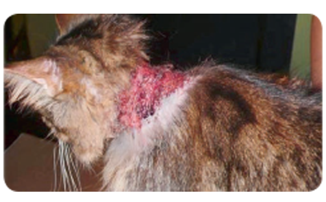
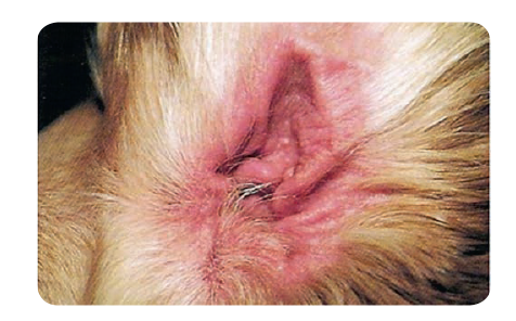
In 14 dogs
First cytology – numerous inflammatory cells – degenerate and non-degenerate neutrophils, chromatin threads from damaged neutrophils, several cocci between chromatin threads and up to 20 yeasts in the field of view.
Second cytology – isolated clusters of neutrophils, up to 10 yeasts in the field of view, no cocci.
Group 2 (30 dogs)
In 21 dogs of the following breeds: French Bulldog, English Bulldog, Boxer, and Pug with multifold facial dermatitis and visible exudate and maceration of the epidermis within the skin fold
First cytology – numerous yeasts – up to 20 in the field of view and isolated cocci, no inflammatory cells
Second cytology – up to 5 yeasts in the field of view, no cocci
In 9 dogs of the above-mentioned breeds showing the same clinical symptoms
First cytology – numerous yeasts (up to 15 in the field of view) and degenerate and non-degenerate neutrophils
Second cytology – no yeasts,
In 5 dogs, no inflammatory cells were observed
In 4 dogs, isolated clusters of non-degenerate neutrophils were observed.
Group 3 (12 cats)
In 8 European shorthair cats with self-inflicted skin erosions on the head and neck and with inflammatory exudate – first cytology – non-degenerate and degenerate neutrophils, no microorganisms
1 cat did not report for follow-up
Second cytology
In 5 cats – second cytology – several inflammatory cells – isolated non-degenerate and degenerate neutrophils
In 2 cats – normal physiology
In 4 cats of the same breed and the same symptoms, first cytology – additional numerous cocci phagocytized by neutrophils, second cytology – isolated neutrophils and individual cocci.
Second cytology – isolated epithelial cells of the granular layer and corneal cells in 3 cats, in all 8 patients – dry changes without exudate on the wound.
According to my findings, Oticurant brings new treatment opportunities, especially whenever the antibiogram suggests that all other methods have been exhausted, e.g. in cases caused by highly resistant staphylococcal strains.
It is also worth using this new product for preventive purposes in patients with diagnosed atopic dermatitis and in dogs that like to bathe in stationary water basins during the swimming season. Oticurant may also prove useful in patients with risk factors of otitis externa – prevention, after all, is better than cure.
As has been shown, Oticurant can be administered to a great variety of canine patients and will no doubt become a permanent element on the shelves of many veterinary practices.
OtiCurant- The effect of an ear powder on clinical signs in canine with otitis externa.
Mikaela Heidrich, DVM.
EVIDENSIA
Djursjukhuset Karlstad
Stallplatsvägen 2
654 65 Karlstad
Sweden
Email mikaela@djursjukhusetkarlstad.se
Conflict of Interest
Omnidea AB provided partial funding for the
research project to EVIDENSIA, Djursjukhuset
Karlstad, Sweden.
ABSTRACT
Background: Antibiotic resistance is a serious and growing problem, in particular when it comes to pathogenic organisms. One of the most common ailments affecting the canine population is otitis externa and the main bacterium implicated in otitis externa is Staphylococcus Pseudintermedius, which has been shown to be resistant to antibiotics. It has also been reported that Pseudintermedius is zoonotic, which may pose public health concerns. It would therefore be desirable to
find a prophylaxis for otitis externa that both the dog and owner finds convenient and easy to use. What we want to investigate in this study is if there is way to inhibit the overgrowth of microorganisms without pharmaceutical compounds and by doing so limit the risk of future ear problems.
Objectives: The hypothesis is by administering a powder in canine ears consisting of an organic acid, absorbents of moisture and fat, and specific sugars, clinical signs of otitis externa will decrease. Thus implicate that there has been an inhibition of microbial overgrowth. Additionally we investigate whether a powder is convenient to use for dogs and their owners.
Methods: 17 privately owned dogs with clear clinical signs of otitis externa of different severity were randomized into two groups and were treated with an ear powder for 14 days. Clinical signs were determined before and after treatment on day one and 14 by two veterinarians.
Results: A significant decrease in overall clinical signs was observed in the study (p<0,05). Out of the 17 dogs participating in the study 76% showed an overall improvement. Foul odor, pruritus, head shaking and excessive accumulation of lipid/wax were significantly reduced (p<0,05). The powder was well tolerated and well accepted by the dogs and their owners. No adverse side effects were observed. All dogs enrolled completed the study.
Conclusions: The use of the powder is a safe and an effective measure to reduce clinical signs of otitis externa. No buildup of powder in the ear canal was reported and all dogs enrolled completed the study. The administration of the powder was well accepted by the dogs and the compliance was 100 percent. This powder may provide analternative therapeutical and prophylactic approach to lower the risk of overgrowth of microorganisms that can cause ear infection.
INTRODUCTION
Ear problems in dogs are a common ailment that affects an estimated 15-20 percent of total canine population.1 Clinical signs of otitis externa are foul odor, pruritus, head shaking, lipid/wax depositions and erythema. Clinicians must consider the underlying mechanism responsible for otitis externa when examining dogs with ear problems. There are primary causes, perpetuating and predisposing factors that will decide the therapy for ear disease. Primary causes of otitis externa could be parasites, hypersensitivity, keratinization disorders and autoimmune diseases. These conditions are responsible for altering the environment in the ear canal to allow for abnormal colonization of microorganisms. Perpetuating factors include bacteria (primarily Staphylococcus spp. and Pseudomonas spp.), yeasts (primarily Malassezia spp.) and pathologic changes, such as glandular hyperplasia, epithelial folds, neoplasia, edema, mineralization, and fibrosis. Predisposing factors are pendulous pinnae, excessive wax production, high humidity, stenosis and hair in the ears. Alterations in normal microflora in the ear and skin may play a role as predisposing factors in allowing the overgrowth of Malassezia organisms. Malassezia Pachydermatis is a common commensal lipophilic yeast of the anal sacs, anus, auditory canal, and skin of dogs. Malassezia may be found on the skin in as many as 50 percent of healthy dogs and is a common etiological agent in otitis externa. It has been reported that Malassezia Pachydermatis can cause nosocomial infection in humans. Factors favoring its growth include abnormal levels of ceruminous lipids, high humidity, and abnormal cell-mediated immunity. Another prerequisite for optimal growth is a pH in the range of five to eight with a marginal to null growth around pH 3. Staphylococcus Pseudintermedius is a common commensal of oral, nasal, and skin flora in healthy dogs, where it can also cause invasive disease. In humans, it is recognized as an invasive zoonotic pathogen and has been isolated from 18% of canine-inflicted wounds. Pseudintermedius species has been shown to be resistant to antibiotics which is a cause of concern to public health. The optimal pH level for growth is between 7 and 7,5. At pH 5 the growth will be inhibited and under pH 4 it will not grow. It has also been reported that Staphylococcus is a biofilm producer at certain pH levels. The capability to produce biofilm is inhibited at pH 3. The concerned microorganisms are sensitive to changes in the physical environment in the ear. Moisture, lipid levels and pH changes can significantly change optimal growth conditions and disturb colonization. Treatment and prophylaxis therapy for otitis externa typical involve careful cleaning of the auditory canal with liquid ear cleaners. By removal of lipid substrates the necessary conditions for growth and reproduction of the organisms are eliminated. There are numerous ear cleaners commercially available containing a variety of ingredients, such as alcohols, organic acids, propylene glycol, various peroxides and detergents all in liquid preparations. However, liquid preparations have some disadvantages as dogs may resist liquids in the ear. The authors wanted to investigate whether a powder containing lactic acid, kaolin, lactose, L-fucose and HMO (Human Milk Oligosaccharides) could have an effect on clinical signs of otitis externa as well as to evaluate the acceptance by dogs when receiving powder in the ears. It has been demonstrated in numerous papers that by lowering pH with organic acids the microbial growth will be inhibited. This effect is at its highest when there is a maximum of dissociated acid. The proportion of dissociated and undissociated acid is equal when the pH is equivalent to the pKa. L-Fucose and HMO are special sugars that in certain cases can connect to lectins on bacteria and by doing so inhibit the adhesion to cells. It has been shown that Fucose and Galactose has an antiadhesive property on P. Aeruginosa in humans.9 Further L-Fucose has been shown to have wound healing properties by modification of dermal fibroblasts thru collagen biosynthesis. HMO may also modulate epithelial and immune cell responses. The authors have no intention to show that the application of a powder is a treatment in the pharmaceutical sense, rather a prophylaxis for ear infection by controlling the microbial overgrowth.
MATERIALS AND METHODS
The study was designed to make the participants their own controls. In the design of the study it was decided that swabbing would not be used as the quantification using swabbing may pose difficulties and uncertainty when it comes to the evaluation of the results. Rather a more broad approached was chosen where clinical signs of otitis externa would serve as a sign of microbial overgrowth. The use of clinical signs of otitis externa is a wellestablished diagnostic tool in the initial phase of an examination. The participants were randomized and divided into two groups each with a dedicated veterinarian. On day one all participants met with a veterinarian at the clinic for an initial examination. Each clinician examined the dogs by ocular exam and graded the clinical sign on a scale from zero to five. Where zero is no clinical sign and five is a severe clinical sign, although not serious enough to be treated with antibiotics or other forms of pharmaceuticals. To be included in the study the patient had to be exhibiting at least one clinical sign of pruritus, head shaking, foul odor, excessive lipid/wax buildup or erythema. Patient with a more severe manifestation of clinical signs with a grade exciding 5 of the clinical sign scale were excluded from participating in the study. After the initial examination the veterinarian showed the participant how to give the powder by administering the first dose. The dose given was adapted to the size of the ear of the specific dog. The participants were given a canister with the powder and a measuring spoon together with a report card. They were asked to give one dose once per day for the following four days and one dose on day nine and 13. They were also asked to make comments on the report card of their observations from day two to day 13. On day 14 the participants came back to the clinic and was once again examined by the same veterinarian that did the first examination. The veterinarian then conducted a final grading of the clinical signs. Twenty privately owned dogs with documented clinical signs for otitis externa was enrolled in the study. The total amount of ears examined was 32. Three participants did not follow the protocol and were therefore excluded from the final report. Of the 17 dogs that completed the study 10 were males and 7 females with an age of 2 to 13 years. Of the 17 dogs 13 had a previous history of ear problems, 10 of them in the last 12 months. The results below are in the case of odor, excessive accumulation of lipid/wax and erythema based on individual ears (n=32).
STATISTICAL ANALYSIS
Two-tailed paired t-test was used to compare results from visit one and two. A level of P < 0.05 was used to indicate a statistically significant difference.
RESULTS
A significant decrease in overall clinical signs was observed in the study (p<0,05). Out of the 17 dogs participating in the study 76% showed an overall improvement. Pruritus and Head Shaking were both significantly better (p<0,05). For 13 dogs out of 17 with pruritus the average clinical sign grade at the initial examination was 2,7 and at the final examination 0,7 and for head shaking it went from 2,7 to 0,4.

Excessive accumulation of lipid/wax was significantly reduced (p<0,05). The average clinical sign grade for 23 ears of 32 went from an average of 3,0 to 1,3. Foul odor was also significantly reduced (p<0,05) where 13 ears had an average clinical grade of 2,3 at the initial grading and 0,8 at the final examination. Of the 32 ears 6 did not have any odor at the beginning or the end of the study. Another 6 ears had no odor in the beginning but all received grade 1 on day 14. At the same time these 6 ears all had gone from grade 3 in pruritus to grade 0 and from 2, 4 and 3 to 0, 0, 1 in shaking head. There was no significant change in the clinical sign grades for erythema although 50 % of the ears improved. 37 % showed a slight worsening. The rest were unchanged. The powder was well tolerated and no adverse side effects were observed. No obstruction of the powder in the ear canal was reported. The dog owners reported that it was easy and convenient to administer the powder, as the dogs did not resist when given the daily dose. All dogs enrolled completed the study.
DISCUSSION
To the authors’ knowledge, there are no published reports on the use of an ear powder in dogs with otitis externa. The primary objectives of this study were to evaluate a powder and it´s effect on clinical signs of otitis externa and the acceptance by the participating dogs in respect to receiving a powder in the ear. All clinical signs except erythema were significantly improved in this study. We cannot explain why erythema alone did not significantly improve as the rest did. It may be that the duration of the study was too short. It could be of interest to look further into this by conducting a study with a longer duration. It is clear that the powder have physical properties that absorb moisture, lipids and lowers pH below 4. The powder is composed of several different components, none of them can be considered an active ingredient alone but the combination obviously have an additive effect on the clinical signs studied and as stipulated an effect on microbial growth.
LACTIC ACID
Lactic acid is natural organic acid that has a quick onset as it dissolves rapidly in water. The pH for the powder dissolved in water is just below 4, which is equivalent to lactic acids pKa of 3,86. This will give the maximum inhibitory effect on microorganisms. Lactic acid is highly hygroscopic as well; giving an additive effect to lactose and kaolin that are the two main absorbents of moisture and lipids in the formulation.
KAOLIN AND LACTOSE
Some lipid dependent Malassezia species have been isolated in canine ears but the most common yeast is M Pachydermatis. For this yeast lipids is not essential for growth although needed for a rapid growth.12 Kaolin and Lactose are two effective absorbent of lipids and moisture keeping available lipids at a minimum. Both ingredients are highly hygroscopic substances that dry out the humid ear canal removing the moisture necessary for Malassezia to function.
L-FUCOSE AND HMO
The scope of the study was to evaluate clinical signs not separate ingredients. Having said that, based on the literature it could be of interest to conduct further studies with the objective to evaluate these sugars role in inhibiting microbial growth.
EXCESSIVE LIPID/WAX DEPOSITS
A surprise finding in the study was the significant lowering of excessive lipid/wax deposits. During the study period the participants were not allowed to clean the ears of their dogs. When considering that a powder was poured into the ear it may be expected that excess powder would accumulate in the ear. This was not the case in this study. Actually the opposite was true as ears were significant cleaner (p<0,05) at the end of the study than on day one. It was considered whether Epithelial Migration (EM) could be the answer to the significant result. EM is the self-cleaning mechanism of the ear canal as well as the tympanic membrane. Although difficult to measure, several studies have tried to evaluate the EM rate. One study report EM rates on the tympanic membrane between 96.4 (±43.1) µm ⁄ day and 225.4 (±128.1) µm ⁄ day in healthy dogs.13 There are other studies reporting similar results. In this study the participants all showed clinical signs of otitis externa. It has been speculated that otitis externa may obstruct the EM.14 Considering that this study only lasted 14 days the EM does not explain why the study population had significant cleaner ears.
PHYSICAL ACTION
Once the ingredients in the powder come in contact with a moist surface it will directly dissolve and cling to the moist surface. When the powder has saturated the surface the rest of the powder will be free flowing and the superfluous powder may fly out. It has been noted ex vivo that the powder initially absorbs moisture and fatty substances and subsequently after the moisture evaporate building flakes that have a tendency to fall off. This may be an explanation to cleaner ears. However this must be studied in a canine population.
COMMENTS FROM DOG OWNERS
On the report cards filled out by dog owners several commented on the ease of using the powder unlike any liquid earlier used. Their dogs did not resist when they applied the powder, which resulted in 100 percent compliance. Some of the participants reported improvement in clinical signs after three to five days.
CONCLUSION
The use of the powder is a safe and an effective measure to reduce clinical signs of otitis externa. The powder was well tolerated and no adverse side effects were observed. No buildup of powder in the ear canal was reported and all dogs enrolled completed the study. The administration of the powder was well accepted by the dogs and the compliance was 100 percent. This powder may provide an alternative therapeutical and prophylactic approach to lower the risk of microbial overgrowth that can cause ear infection.
ACKNOWLEDGEMENT
The author likes to thank all colleagues that participated in the study along with Omnidea AB, Stockholm, Sweden that supplied the powder for the study.
EFERENCES
- Scott DW, Miller WM, Griffin CE.
Muller & Kirk’s Small Animal
Dermatology. 5th ed. Philadelphia, PA:
WB Sunders; 1995:351-357, 970-987.
- 2. Plant JD, Rosenkrantz WS, Griffin CE.
Factors associated with and
prevalence of high Malassezia
pachydermatis numbers on dog skin. J
Am Vet Med Assoc. 1992;201:879- 882.
- Prevalence of Malassezia
pachydermatis in dogs with suspected
Malassezia dermatitis or otitis in
Slovakia. Eva Čonková, Edina
Sesztáková, Ľubomír Páleník, Peter
Smrčo, Ján Bílek. ACTA VET. BRNO
2011, 80: 249–254;
doi:10.2754/avb201180030249
- 4. Evaluation of the effect of pH on in
vitro growth of Malassezia
pachydermatis
Jennifer L. Matousek, Karen L.
Campbell, Ibulaimu Kakoma, Philip F.
Solter, and David J. Schaeffer. Can J
Vet Res. 2003 January; 67(1): 56–59.
- Clinical Isolates of Staphylococcus
intermedius Masquerading as
Methicillin-Resistant Staphylococcus
aureus. Sudha Pottumarthy1, Jeffrey
- Schapiro1, Jennifer L. Prentice1,
Yolanda B. Houze1, Susan R.
Swanzy1, Ferric C. Fang1,2 and Brad
- Cookson1,2,*J. Clin. Microbiol.
December 2004 vol. 42 no. 12 5881-
5884
- Antibiotic Resistance Profiles of
Staphylococcus pseudintermedius
Isolates from Canine Patients in
Korea. Yoon, Jang Won, Ki-Jong Lee,
So-Young Lee, Min-Joo Chae, JaeKeun Park, Jong-Hyun Yoo, and HeeMyung Park. J. Microbiol. Biotechnol.
(2010), 20(12), 1764–1768 doi:
10.4014/jmb.1001.01011 First
published online 7 October 2010
- A Microtiter plate assay for
Staphylococcus aureus biofilm
quantification at various pH levels and
hydrogen peroxide supplementation.
Tarek Zmantar, Bochra Kouidhi,
Hanene Miladi, Kacem Mahdouani,
Amina Bakhrouf. NEW
MICROBIOLOGICA, 33, 137-145, 2010
- Staphylococcal biofilm formation as
affected by type acidulant.
Antonia Nostro, LuignaCellini,
Giovanna Ginestra, Manuela D´Arrigo,
Mara Di Giulio, Andreana Marino,
Anna Rita Blanco, Angelo Favaloro,
Giuseppe Bisignano. © 2013 APMIS.
Published by John Wiley & Sons Ltd
- Inhalation with Fucose and Galactose
for Treatment of Pseudomonas
Aeruginosa in Cystic Fibrosis Patients
Hans-Peter Hauber Maria Schulz
Almuth Pforte Dietrich Mack Peter
Zabel Udo Schumacher
Int. J. Med. Sci. 2008 5(6):371-376
- Role of sugars in surface microbe–host interactions and immune reaction modulation
David H. Lloyd, Jacqueline Viac, Dirk
Werling, Christophe A. Rème, Hugues
Gatto
© 2007 The Authors. Journal
compilation © 2007 ESVD and ACVD.
18; 197–204
- Human Milk Oligosaccharides: Every
Baby needs a Sugar Mama
Lars Bode
The Author 2012. Published by Oxford
University Press.
- 12. Atypical Lipid-Dependent Malassezia
Species Isolated from Dogs with Otitis
Externa M.J. Crespo, M.L. Abarca, F.J.
Cabanes. JOURNAL OF CLINICAL
MICROBIOLOGY, June 2000, p.
2383–2385
- Epithelial migration on the canine
tympanic membrane. Natalie E.
Tabacca, Lynette K. Cole, Andrew
Hillier, Pä ivi J. Rajala-Schultz
Veterinary Dermatology DOI:
10.1111/j.1365-3164.2011.00982.x
- 14. Logas DB. Diseases of the ear canal.
Veterinary Clinics of North
AmericaSmallAnimalPractice1994;24:9
05–19.
elves of many veterinary practices.
UrinoVet Dog - Result of clinical trial of the medication UrinoVet Dog
 Clinical trial of the medication Urinovet Dog produced by Vet Planet Ltd. was conducted in the Veterinary Policlinic and the Clinical Diagnostics Faculty of the Veterinary Medicine Department at the Warmian-Masurian University in Olsztyn. The drug was delivered directly by the manufacturer Vet Planet , Staszica Street 20 A, 05-092 Lomianki.
Material and methods
The trial, lasting from January 1st 2010 to March 31st 2010, was conducted among 44 dogs of the different sex, age and race. It was divided into two phases: in the first one, based on the clinical examination and the additional tests, ill animals were diagnosed. In the second phase the clinical efficacy of the medication Urinovet Dog was assessed. All the animals had symptoms of the lower urinary tract disorders: pollakiuria, urinary urgency, stranguria, dysuria, vocalization during urination, haematuria, periodically smelly urine, urination in the form of droplets, urinary incontinence.
The animals were divided into four groups, depending on the age and sex:
I- 10 young and middle-aged male dogs (age 2- 6 years)
II- 10 young and middle-aged female dogs (age 2.5- 7 years)
III- 14 old male dogs (8- 12 years)
IV- 10 old female dogs (9- 12 years)
The young and middle-aged animals (groups I and II) were neither castrated nor sterilized. In the III group 4 male dogs were castrated, in the IV group 5 female dogs were sterilized. The common antiparasite preventive measure and the prophylactic vaccination were carried out. Most of the animals (32) were fed with commercial fodder of different companies, dry as well
as wet food; the rest (12) was given home diet food, based mainly on the poultry meat, rice or noodles. They had a constant access to water. They were the only animals present at home. All the dogs had the clinical examinations, blood cell count tests, serum biochemical tests and the general urinalysis along with the examination of the urinary sediment. Also
ultrasonography of the urinary tract was performed.
It was concluded based on the results of the above mentioned tests, that:
Gr I- 8 male dogs – acute cystitis (I a)
2 dogs- recurrent chronic cystitis (I b)Gr II- 5 female dogs – acute cystitis (II a)
4 female dogs- recurrent chronic cystitis (II b)
1 female dog- struvite urolithiasis (phosphate-ammonium-magnesium) (II c)- status
after catheterization and cystolithotomy
Gr III- 8 male dogs- recurrent chronic cystitis (III a)
2 male dogs- struvite urolithiasis (phosphate-ammonium-magnesium) (III b)
3 male dogs- struvite urolithiasis (phosphate-ammonium-magnesium) (III c)- status
after catheterization and cystolithotomy
1 male dog- oxalate urolithiasis (III d)
Gr IV- 7 female dogs- chronic cystitis (IV a)
2 female dogs- struvite urolithiasis (phosphate-ammonium-magnesium) (IV b)
1female dog- oxalate urolithiasis (IV c)- status after catheterization and cystolithotomy
The medication UrinoVet Dog was the only one and the basic drug given to ill animals. It was administered during 3 weeks (21 days) in quantity recommended by the manufacturer- 1 tablet/ 5 kg – in case of acute cystitis (groups Ia, IIa); 1 tablet/ 5 kg during 6 weeks (42 days) – in case of chronic cystitis and urolithiasis ( groups Ib, IIb, IIIa, IIIb, IIId, IVa, IVb). The dogs
that underwent surgical treatment were given the drug in dose of 2 tablets/ 5 kg during 5 days, afterwards in dose of 1 tablet/ 5 kg during 6 weeks (42 days) (groups IIc, IIIc, IVc). The preparation was mixed with the wet food or given directly to the mouth. Health status of all the dogs, taking part in the trial, was monitored in the clinical checkup weekly.
The results of the trial
Gr I –Ia- evident improvement (EI) just after 14 days of using the preparation in all 8 dogs
Ib- evident improvement (EI) in 1 dog after 21 days, in 1 dog-slight improvement (SI)
GrII- IIa- evident improvement (EI) in 4 female dogs, slight improvement (SI) in 1 female dog
IIb- evident improvement (EI) in 3 female dogs, slight improvement (SI) in 1 female dog
IIc- evident improvement (EI) after 5 days of use of the drug in higher dose (2 tablets/5kg) for 5 days; evident improvement (EI) after the next 6 weeks of using the drug (dose 1 tablet/ 5 kg). The female dog has been still given the medicament in the maintenance dose (1tablet/ 10 kg)
Gr III- IIIa- evident improvement (EI) in 5 male dogs, slight improvement (SI) in 1 male dog, lack of improvement (LI) in 2 male dogsIIIb- evident improvement (EI) in 1 male dog, slight improvement (SI) in 1 male dog IIIc- evident improvement (EI) in 2 male dogs (they have been still given the drug in the maintenance dose 1 tablet/ 10 kg), lack of improvement (LI) in 1 male dog
IIId- slight improvement (SI) in 1 male dog Gr IV- IVa- evident improvement (EI) in 5 female dogs, slight improvement (SI) in 1 female dog, lack of improvement (LI) in 1 female dog
IVb- evident improvement (EI) just after 5 days in 1 female dog, slight improvement (SI) in 1 female dog after 5 days; UrinoVet Dog was given 7 days longer in dose of 2 tablets/5kg – evident improvement (EI). Both female dogs have been still given the drug in dose of 1 tablet/ 10 kg- lack of the clinical symptoms of the urinary tract disease
IVc- evident improvement (EI) in 1 female dog (it has been still given the drug in the
maintenance dose of 1 tablet/ 10 kg)
Evident Improvement (EI)- lack of any clinical symptoms of the lower urinary tract
Slight Improvement (SI)- urinary urgency, dysuria, periodic pollakiuria, slight haematuria (1-
2 days), periodically smelly urine
Lack of Improvement (LI)- pollakiuria, urinary urgency, stranguria, dysuria, vocalization
during urination, haematuria, periodically smelly urine, urination in the form of droplets,
urinary incontinence.
Conclusion
The preparation, mixed with wet fodder or given directly to the mouth, was readily eaten by the animals. Side and undesirable effects were not noted during the clinical trial. The medication slightly increased the water intake by the dogs, what gave a beneficial effect, boosting its activity by increasing diuresis and removing toxic and noxious substances from the lower urinary tract.
The drug occurred to be very effective for 13 animals having acute cystitis- evident improvement (EI) was noticed in 12 animals, what amounts to 92.3 %. Slight improvement was noted in 1 female dog (7.7%). In the group of the animals with chronic cystitis evident improvement (EI) was observed in 14 dogs of 21 examined (66.7%), slight improvement (SI) in 4 animals (19%) and lack of improvement in 3 animals (14.3%). In the dogs diagnosed with urolithiasis the drug occurred to be effective in 40%- evident improvement (EI) was noted in 2 dogs of 5 examined. In 3 animals slight improvement (SI) was observed- 60%, but after increasing dose to 2 tablets/ 5 kg evident improvement was noted on the next 7 days. Cystolithotomy was performed in 5 animals having urolithiasis. Earlier the animals had been catheterized, due to the urethra obstruction. They were given UrinoVet Dog in higher dose (2 tablets/ 5 kg for 5 days). In 4 of 5 animals evident improvement (EI) was noted- 80%. In 1 dog (20%) lack of improvement (LI) was observed. All the animals that underwent the surgical procedures have been still given the drug in the maintenance dose (1 tablet/ 10 kg) without recurrence of the clinical symptoms of the urinary tract diseases.
Summarising, one can note, that the preparation UrinoVet Dog produced by Vet Planet may be effective in treatment of the canine urinary tract diseases. The medication does not cause side and undesirable effects in dose of 1 tablet/ 5 kg, as well as in dose increased by 100%, independently from the length of the application. It may be used in the treatment and the prophylaxis of the canine urinary tract diseases.
VetoSkin - Pilot clinical trial on the VetoSkin® preparation in dogs with atopic dermatitis
 Abstract
The aim of this trial was to evaluate the clinical effect of VetoSkin® supplementation in dogs with atopic dermatitis fed the same diet. The control group consisted of 10 dogs selected to meet the same criteria as the trial group. All patients were given one capsule of VetoSkin® per 10 kg body weight. Clinical examination was made on the day 0, 30, 60 and 90. The CADESI 03 and PVAS (Pruritus Visual Analog Scale) scoring systems were used to evaluate the results during the three month trial.
Key words
atopic dermatitis, atopy, essential fatty acid supplementation, eicosapentaenoic acid, gamma-linolenic acid.
Essential unsaturated fatty acids (n3 and n6) have been widely used in the treatment of atopic dermatitis for more than 25 years. Their clinical signifcance has so far been proved in about twenty studies (1-3). Currently, essential unsaturated fatty acids are widely used in the treatment of atopic dermatitis in dogs (7, 9). They inhibit the synthesis of LBT4 by re-balancing the hydrolipid barrier on the whole surface of the dog’s skin. Polyunsaturated fatty acids are components of cell membranes their oxidation contributes to the formation of prostaglandins and leukotrienes, two eicosanoids which take part in the development of inflammatory conditions. Both the eicosapentaenoic and gamma-linolenic acids (omega-3 and omega-6 respectively) participate in the metabolism of arachidonic acid by competing with the same enzymes, and thus reduce the production of inflammatory eicosanoids (e.g. PGE2, PGI1, LTB4). This, in turn, favours the production of anti-inflammatory eicosanoids such as PGE1, PGE3 or LT5. Eicosapentaenoic acid can be found in oil obtained from some fsh and the highest concentration of gamma-linolenic acid is in oils from evening primrose and borage (4, 5, 7). Preparations containing essential unsaturated fatty acids combined with other substances such as vitamins, minerals or cofactors have been a relatively new area and further experiments are needed to evaluate their effectiveness (7, 8). Many producers believe that the right combination of a few substances will maximize their effciency (7). So far, however, there are no studies concerning those types of products. No studies which would confrm that effciency have yet been presented. In one study involving a double-blind trial the product containing polyunsaturated fatty acids and cofactors proved less effective than a product without cofactors (10). However, further research is needed. The analysed Vetoskin preparation contains omega-3 and omega-6 acids as well as B vitamins, biotin and zinc. B vitamins are constantly synthetized by the intestinal flora but as they are water-soluble, they are not stored in the body. Therefore, they have to be continuously supplied. Defciency in group-B vitamins may lead to skin conditions. Such conditions, however, are not very specifc and other diseases should be taken into consideration in the process of differential diagnosis. Clinical symptoms of a B-group vitamin defciency include dull hair, dry seborrhea and hair loss on some facial areas. Vitamin B (pyridoxine) takes part in the metabolism of many nutrients relating to the normal functioning of hair and skin such as the transformation of linoleic acid into arachidonic acid, methionine into cysteine and tryptophan into niacin (vit. PP). It also participates in the synthesis of picolinic acid, crucial for zinc to penetrate the intestinal mucosa (7, 8). Zinc is an integral part of many metalloenzymes which participate in regulating the metabolism. It is an important cofactor for the RNA and DNA polymerases and particularly important for fast dividing cells such as those of the epidermis. Zinc is essential for the biosynthesis of fatty acids and takes part in the metabolism of vitamin A. Zinc plays a crucial role in ensuring the normal functions of the immune system and is also present in inflammatory reactions. The dermatological symptoms of zinc defciency include: slow healing of wounds, local erythema, patches of hair loss, crust and desquamation. The above-mentioned lesions usually occur in places prone to injuries, such as skin-membrane connections, distal parts of the body and fngertips. Fur becomes dull and bacterial and fungal infections occur. However, diet-related zinc defciency is infrequent (7). Biotin (vitamins H and B7) is a
co-enzyme of a few various enzymes. It is an essential component of biotin-dependent carboxylases. Carboxylases are enzymes which are vital for many important biochemical reactions, for example in the process of the formation of glucose (gluconeogenesis), the synthesis of fatty acids or the cycle of citric acid. Biotin supports the normal functions of the thyroid gland and contributes to the normal functioning of the skin and hair. Symptoms of biotin defciency include dermatitis, urticaria and hair loss. Other systemic manifestations may include an elevated level of cholesterol and inflammatory lesions in the bowels. As biotin can be synthetized by the intestinal flora, its deficiency occurs very rarely and is usually caused by other than nutritional factors. Biotin defciency happens during a prolonged antibiotic therapy which is often prescribed in the treatment of post-atopic dermatitis bacterial infections (phlegmon) (1, 7, 9).
Material and methods
The study was carried out on 20 dogs as the study group (group I) and 10 dogs as the control group (group II). All dogs had atopic dermatitis. The diagnosis of atopic dermatitis in the animals from both groups was made based on the diagnostic criteria of Claude Favrot (12, 13) and intradermic tests (Agroskin RTU 20; Agrolabo). Additionally, before carrying out the intradermic test, all dogs were subject to rapid diagnostic tests to assess the total number of IgE antibodies (VetExpert). The tests gave positive results in all cases. The intradermic tests were conducted at least 3 months before the animals were qualifed for the study. All the dogs were found sensitive to many all-year allergens. The dogs with seasonal allergies were not included in the study. In all dogs from both groups the symptoms of atopic dermatitis of varied intensity had been present for the whole year. The study group consisted of dogs aged 2-6 years (the average of 4.2 years), 10 females, including 4 spayed, and 10 males, including 5 castrated ones. The dogs represented different breeds (5 Labradors, 5 mongrels, 3 West-Highland White Terriers, 3 Beagles, 3 German Shepherds and 1 Dachshund) and their body weight ranged from 10 to 30 kg. The control group consisted of 10 dogs aged 3-6 (the average of 5 years), 5 females, including 4 spayed ones and 5 males, including 3 castrated ones. The dogs represented different breeds, 2 Labradors, 3 mongrels, 2 West-highland white terriers and 3 German shepherds of body weight ranging from 10 to 40 kg.
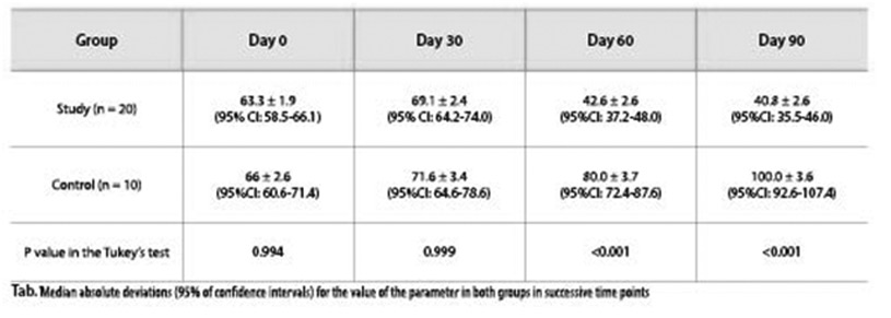


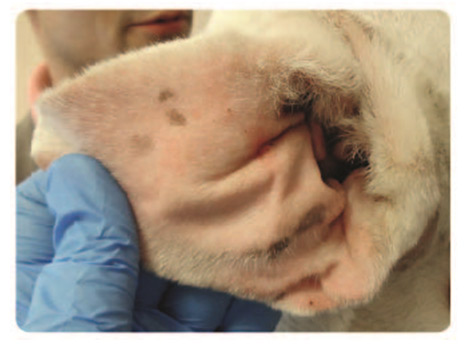
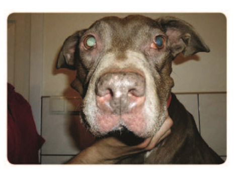

Food allergies were excluded in all dogs by using hydrolyzed elimination diet for at least 12 weeks. Throughout the study all dogs from both the study and the control groups followed the same diet (Hypoalergenic®Royal Canin). The dogs did not undergo allergen immunotherapies. During the three-month study the dogs did not receive anti-inflammatory drugs, glucocorticoids, antibiotics, antihistamines or cyclosporine. The animals from the study group received VetoSkin® containing B-group vitamins (B1, B2, B6 and B12), NNKT-Omega-3/Omega-6, biotin and zinc. The product was administered to the dogs by their owners at home for 12 successive weeks (90 days/3 months) in March, April and May. The dosage of the product was determined based on the animal’s body weight and was as follows: 1 capsule of VetoSkin® for every 10 kg of body weight. The capsules could be administered in whole or, as they are twist-off type, it was also possible to only give the content. The dogs from the control group did not receive VetoSkin®. All dogs underwent dermatological examinations four times on the following days: 0, 30th, 60th and 90th. The intensifcation of lesions was assessed by using the CADESI 03 system. The levels of erythema, lichen, skin abrasion and hair loss caused by self-mutilation in 62 areas of the body were analyzed. The following point-scale was used to assess the lesions: 0 – no lesions, 1 – mild lesions, 2, 3 – moderate lesions and 4, 5 – considerable lesions. All dogs were in remission from the disease and mild to moderate intensity of the symptoms of atopic dermatitis was observed. On the 0 day the dogs scored up to 84 points in the CADESI system. Additionally, the intensity of pruritus in the PVAS (Pruritus Visual Analog Scale) 5-stage scale was measured. The criteria defning pruritus used in the study were according to Marselli et al.: 0 points – no pruritus, 1 point – mild pruritus, the animal scratches itself for less than 10% of the observation time, 2 points mild to moderate pruritus, 30% of time spent on scratching, 3 points moderate pruritus, 30-50% of time spent on scratching, 4 – moderate to considerable, 50-75% of time, including at night and 5 – considerable pruritus when the animal scratches
Ryc. 3. Rumień okolicy twarzowej u psa z AZS
Fig. 3. Erythema on the facial area of a dog with AD
Ryc. 4. Niewielki rumień skóry okolicy przestrzeni międzypalcowych u WHWT z atopią o łagodnym przebiegu
Fig. 4. Small erythema in the interdigital area of a WHWT with mild atopic dermatitis itself for more than 75% of time, including at night and while eating. The assessment was done by the owners on the following days of the study: 0, 30th, 60th and 90th and the results were marked on the scale of pruritus intensity. Statistical analysisThe quantitative variable was presented as an average standard deviation. Additionally, a 95% confdence interval (95% CI) was calculated for average group means. The statistical analysis was conducted using a two-way repeated measures analysis of variance (group as the random factor and time as the non-random factor). As sphericity was assumed (the Mauchley test p = 0.206), one-dimensional tests were used. The Tukey’s test was used for uneven group numbers in the post-hoc analysis. The result was considered statistically signifcant when the two-tailed p value was less than 0.05. The statistical analysis was carried out by Michał Czopowicz, D.V.M. PhD, from the Laboratory of Veterinary Epidemiology and Economics of the Department of Veterinary Medicine of the Warsaw University of Life Sciences in Warsaw, Poland. Statistically signifcant differences were observed for inter-group comparison (p < 0.001), inter-time points comparison (p < 0.001) and for the interaction of group and time (p < 0.001). The value of the measured parameter changed over time in both groups and the direction of those changes was divergent. In the study group the CADESI value remained the same between day 0 and 30 (p = 0.248) but was statistically signifcantly lower between the 30th and 60th days (p < 0.001) and remained constant between the 60th and 90th days (p = 0.998). Statistically signifcant decrease in the CADESI value happened between the 30th and 60th days. In the control group the CADESI value systematically increased and on the 60th day was statistically signifcantly higher (p = 0.016) compared with day 0, and on the 90th day it was statistically signifcantly higher than on days 0, 30 and even on the 60th day (p < 0.001). The CADESI value was different between the groups. A statistically signifcant difference between the study and control groups occurred on the 60th (p < 0.001) and 90th days (p < 0.001). There was no statistically signifcant difference between the groups on days 0 (p = 0.994) and 30 (p = 0.999). It is good because it means that the groups were comparable at the very beginning of the study.
Results of the study and discussion
At the beginning of the study in dogs from both the study and control groups mild to moderate symptoms of atopic dermatitis were observed. On day 0 the CADESI value in group I ranged from 53 to 84 points (the average of 62) and in group II it was from 49 to 79 points (the average of 65). On the 30th day the CADESI value in group I was from 63 to 83 points (average 70) and in group II – from 49 to 90 (average 75). On the 60th day of the study the CADESI value for group I ranged from 34 to 50 points (average 42) and in group II – from 32 to 130 (average 80). On the 90th day of the study the CADESI value for group I was from 21 to 49 points (average 39) and in group II – from 42 to 120 (average 100). The intensity of pruritus according to PVAS (Pruritus Visual Analog Scale) was also assessed four times. On day 0 in 9 dogs from the study group (45%) mild pruritus was observed, in 8 dogs (40%) it ranged from mild to moderate and was moderate in 3 dogs (15%). On day 0 mild pruritus was observed in 4 dogs (40%), mild to moderate – in 3 dogs (30%) and moderate – in 3 dogs (30%). On the 30th day of the study in 7 dogs from group I (35%) mild pruritus was observed, in 9 dogs (45%) it ranged from mild to moderate and it was moderate in 4 dogs (20%). In group II mild to moderate pruritus was reported in 7 dogs (70%) and in the case of 3 dogs (30%) it was moderate. On the 60th day in 10 dogs from the study group (50%) a mild intensity of pruritus was observed, mild to moderate in 9 dogs (45%) and moderate in 1 case (5%). In the control group the intensity of pruritus was mild to moderate in 5 dogs (50%), moderate in 2 dogs (20%) and moderate to considerable in 3 dogs (30%). On the 90th day of the study in 12 dogs from the study group (60%) the intensity of pruritus was mild and it ranged from mild to moderate in the remaining 8 dogs (40%). On the same day in the control group moderate pruritus was reported in 4 dogs (40%) and in 6 dogs (60%) it ranged from moderate to considerable. The results of the study indicate that after 2 months of constant administration of the VetoSkin® preparation the clinical condition of dogs with atopic dermatitis improved and remained stable for the following 4 weeks. In the control group, where the preparation was not administered, the clinical condition worsened with time. Similar results were obtained in studies conducted earlier (1, 2, 5, 7). In one of the studies it was observed that in young dogs with recently developed atopic dermatitis the response of the body was significantly better after two months. In this study the improvement was observed in both younger (2/3 years) and older (5 years) dogs (2-4). In the study concerning the intensity of pruritus according to PVAS (Pruritus Visual Analog Scale) a considerable reduction in the intensity of pruritus was observed in the study group after 60 and 90 days. In the dogs from the control group the intensity of pruritus increased gradually with time and was followed by complications such as phlegmon which after the study had finished required a treatment with glucocorticoids and antibiotics. In none of the dogs from the study group the side effects mentioned in the available literature and including diarrhea and pancreatitis were noticed (7). The results obtained from the study indicate that VetoSkin® containing polyunsaturated fatty acids, B-group vitamins, zinc and biotin may be used in dogs with atopic dermatitis to alleviate the dermatological symptoms and the intensity of pruritus.